
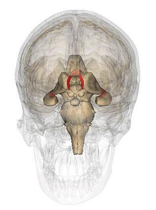
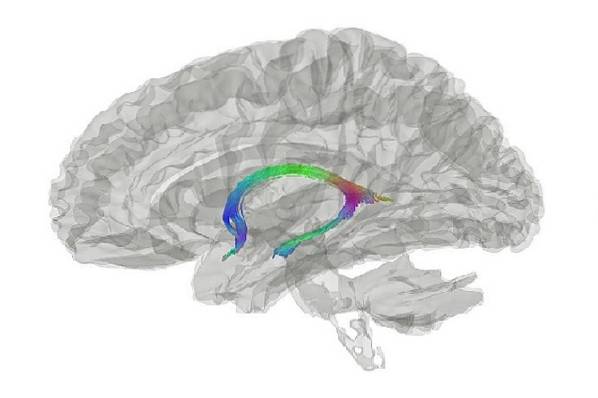
The fornix, Trine of the brain, vault of the four pillars or cul-de-sac, is a brain region formed by a series of nerve bundles. This structure is C-shaped and its main function is to transmit signals. Specifically, it connects the hippocampus with the hypothalamus, and the right hemisphere with the left hemisphere..
The fornix is full of myelinic fibers, that is, of white matter, it is found just below the corpus callosum, and some authors consider it as a part of the limbic system of the brain. Likewise, certain investigations have shown that the relationship of this structure with the hippocampus could play an important role in memory processes.

At present, various investigations have shown that the most important efferent pathway of the hippocampus is the one that connects it with the fornix. Thus, although the hippocampus has many other connections, the most prevalent seems to be the one that relates it to the trigone of the brain..
For this reason, it is theorized that the fornix could be a highly relevant structure that gives rise to many of the functions that the hippocampus develops.
Article index
The cerebral fornix constitutes a bundle of highly myelinated fibers of the telencephalon. The fibers of this region of the brain project from the hippocampus to the hypothalamus, thus connecting the two structures..
Certain authorities consider the fornix as part of the limbic system, although its involvement in this type of brain function is still little studied today.

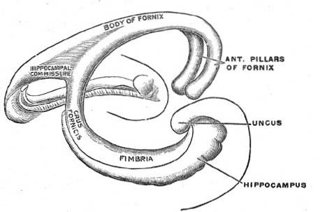
The fornix is an arched “C” -shaped structure just below the corpus callosum. It contains large amounts of white matter, which is why it is considered a communication structure.
Specifically, the fornix seems to play a highly relevant role in memory processes. Many authors think that this structure is vital for the performance of normal cognitive functioning.

The fornix is a small region of the brain. It is located in the telencephalon, just below the corpus callosum. Likewise, the hippocampus is located inferiorly and laterally to the fornix and the amygdala is located between both structures..
The fornix is also known as the trigone or four-pillar vault because it has two anterior projections and two posterior projections. The latter are also known as pillars or columns.
Being a region that contains only white matter, that is, axons of neurons but not bodies of neurons, the fornix is a structure that only performs communication activities between different brain regions.

In this sense, the fornix is a fibrous structure that participates in the union of all those elements of the limbic system, unifying the structures of the right hemisphere with the structures of the left hemisphere..
Thus, this brain region is responsible for connecting the anterior cortical areas with the contralateral posterior cortical areas. That is to say, it allows to cross the information of the different brain regions.
More specifically, the anterior columns of the fornix communicate directly with the posterior nuclei of the hypothalamus, which are known as the mammillary bodies..
Instead, the posterior columns of the fornix establish a connection with the tonsil body (nuclei of the telencephalon that are arranged behind and below the hippocampus).
Thus, in general, the fornix is a brain structure that connects the mammillary bodies with the tonsil nuclei.
Apart from this main connection, the fornix relates more brain regions. The lower part of the structure continues by fibers exiting the hippocampus, thus constituting the hippocampal fimbriae. These fibers form an extension of the posterior columns of the fornix.
Likewise, the mammillary bodies do not only communicate with the fornix, but also establish communication with the anterior thalamic nuclei through the thalamic mammillary fasciculus. Finally, the thalamus communicates directly with the cortex of the frontal lobe through the tenth area of Brodmann.
The main function of the fornix seems to be related to cognitive processes, especially memory function.
The involvement of the fornix in such activities was discovered through surgical trauma, which demonstrated that a disconnection in the fornix involved the appearance of significant cognitive alterations.
In this sense, it is currently argued that the fornix is a fundamental brain structure for the normal cognitive functioning of people.
Likewise, this region could play a very important role in the formation of memory as it is involved in the Papez circuit, a set of nerve structures in the brain that are part of the limbic system..
In summary, the fornix seems to be a very important brain structure in the performance of cognitive activities, since it is responsible for communicating and relating the regions of the brain that perform such actions.
Today it is well established that fornix damage or disease mainly causes cognitive deficits. More specifically, the injury to this brain structure usually generates the experience of retrograde amnesia in the person.
This fact reinforces the data obtained about the activity and functions of the fornix and, at the same time, highlights the alterations that certain diseases can generate.
There are many pathologies that can damage the fornix. However, this does not mean that they always do it or that this brain structure always presents the same lesions and generates the same symptoms..
First, midline tumors or herpes simplex encephalitis can affect the fornix, thus causing certain cognitive failures and / or memory loss..
On the other hand, pathologies or inflammatory conditions, such as multiple sclerosis, can alter the functioning of the fornix and illustrate its importance in global cognitive functioning, generating a generalized dysfunction of cognitive abilities..
The limbic system constitutes a set of brain structures that are responsible for regulating physiological responses to certain stimuli. This system regulates human instincts and actively participates in the performance of activities such as involuntary memory, hunger, attention, sexual instincts, emotions, personality or behavior..

The structures that make up this important brain system are: the thalamus, hypothalamus, hippocampus, amygdala, corpus callosum, midbrain, and septal nuclei.
In this way, the fornix does not constitute a brain region that is part of the limbic system, however, there are many studies that show a close relationship between fornix and the limbic system.
In general, the fornix appears to be related to the limbic system by its location. In fact, the different structures that make up this system surround the fornix, so it is within the circuit that makes up the limbic system..
In more detail, the fornix plays a major role in connecting different regions of the limbic system, such as the thalamic nuclei, the hippocampus, and the tonsillar bodies..
Likewise, it also seems to be one of the main areas of association of the septal nuclei of the brain, transmitting afferent fibers to these structures..
In this way, the fornix is not a main structure of the limbic system but it does play an important role in its functioning. It is an area of association that allows to connect the structures of the limbic system and, therefore, gives rise to its activity.
The element of greatest scientific interest about fornix is its relationship with cognitive impairment. Different studies have examined the role of this brain structure in cognitive pathologies and some research has shown that fornix could predict cognitive impairment.
In this sense, the fornix reveals how not only lesions in the hippocampus (brain structure of memory par excellence) can explain cognitive deterioration, but there are other regions of the brain involved.
In fact, some authors suggest that changes in the structure and function of the fornix could predict in a more detailed way the cognitive decline experienced by healthy people (without dementia) during old age.
Specifically, a study published in the journal Journal of the American Medical Association - Neurology (JAMA-Neurol) identified fornix as the brain structure whose volume loss best predicts the future of cognitive decline among healthy elderly.
The study examined 102 people with an average age of 73 years who underwent clinical evaluations accompanied by magnetic resonance studies.
Although such hypotheses still require further testing, the implication of the fornix in cognitive impairment could be of great relevance, since it could allow a greater understanding of the ins and outs of the continuum from normal cognitive state to dementia.


Yet No Comments