Gardnerella vaginalis It belongs to the Bifidobacteriaceae family within the Bifidobacteriales order of Actinobacteria. This species is characterized because it can behave as Gram positive or Gram negative, depending on the strain and culture medium..
It is facultative anaerobic, producing mainly acetic acid in the fermentation process. It is pleomorphic (it has two structural shapes), and it can be shaped like a bacillus or a coconut (rounded).
The rounded (coccobacilli) and elongated (bacilli) forms can occur in the same strain at different stages of development. Likewise, the type of staining they present (Gram positive or negative) may be affected..
When it was first described, it was placed in the genus Haemophilus. Subsequently, the morphological and functional differences of the species were evidenced. It was located in the genus Gardnerella which is made up of a single species.
Article index
G. vaginalis is facultative anaerobic, although some strains may be obligate anaerobic.
The main product of the fermentation of sugars is acetic acid. However, some strains can produce lactic, formic, or succinic acid. No gases are produced in the fermentation process.
These bacteria can ferment different types of sugars such as dextrin, glucose, maltose and starch.
To recognize this species, the most important characteristics are the hydrolysis of starch and hippurate (aromatic organic compound). Likewise, they generate hemolysis in the presence of human blood, but not in sheep blood..
G. vaginalis it is considered the main causative agent of vaginal bacteriosis. The species is part of the bacterial microflora of the vagina, but can become virulent.
Vaginal bacteriosis is associated with the occurrence of an imbalance of the microbiota in the vagina. Thus, the lactobacilli that produce large amounts of hydrogen peroxide are replaced by anaerobic bacteria.
The species G. vaginalis it inhibits the growth of lactobacillus and the pH of the vagina can increase to values close to 7. The bacterium has the ability to degrade the mucins that are secreted in the epithelial cells of the vagina.
The most obvious symptoms of vaginal bacteriosis are the production of a white or yellowish discharge and a bad smell. Itching and redness may also occur..
The most common forms of infection are unprotected sex and having multiple sexual partners. It is also common to get the disease from sharing sex toys or using the IUD (intrauterine device)..
The most common treatments are the use of antibiotics such as metronidazole and clindamycin..
The optimum temperature for the development of the bacteria ranges from 35 - 37 ° C although they can develop from 25 - 42 ° C. The pH range is 6 - 6.5, but some growth may occur at pH 4.5.
Colonies are not hemolytic in sheep blood. They produce hemolysis growing in human and rabbit blood.
They are considered "fastidious" bacteria, since they require certain specific nutrients for their growth in culture media. Among these we have the presence of biotin, folic acid, thiamine, riboflavin and purines / pyramids..
It has been observed that in the presence of fermentable carbohydrates and peptones, the growth of the bacteria in the medium is accelerated.
The size of the genome in G. vaginalis It is 1,490-1,700 base pairs, with a GC content ranging from 41-43% among the different strains. The core genome (genes shared by all strains) is only 716 genes. In such a way, that only 27% of the genome is common to all the studied strains of the species..
In molecular studies carried out on different strains, it has been determined that at least four different groups are present. These groups have a different genome size and GC relationship to each other.
The species was first isolated in 1953 by Leopold. This author obtained the bacteria from the genitourinary system of men.
The isolate corresponded to a bacterium that behaved as Gram negative, it was immobile and without the presence of a capsule. This first culture was made on blood agar at a temperature of 37 ° C.
Leopold considered that the species was akin to the genus Haemophilus. Later, Gardner and Dukes in 1955 identify it as Haemophilus vaginalis, due to its Gram negative stain and bacillus shape. In addition, they considered that it was the cause of a characteristic vaginal discharge.
However, by continuing the study of the species it was determined that it did not require for its development some elements necessary for the growth of the species of Haemophilus. On the other hand, the bacteria showed a tendency to retain the violet crystal coloration in the Gram stain..
These characteristics indicated that the species was more related to the genus Corynobacterium, which is a Gram positive group of Actinobacteria. For this reason, in 1963 Zinnemann and Turner identified it as Corynobacterium vaginale.
In the 80s of the 20th century, various studies were carried out with biochemical and molecular techniques and observations with the transmission electron microscope. Greenwood and Picket determine that there was no genus with the characteristics of this species.
The authors propose a new genre called Gardnerella in honor of Gardner, which is monospecific (with only one species). They indicate that the bacteria of the genus are Gram negative to variable, rod-shaped and have a laminated cell wall..
Currently the genus is located in the Bifidobacteriaceae family of the Bifidobacteriales order of the Actinobacteria. Recent molecular studies indicate that the species forms a clade with species of the genus Bifidobacterium (B. coryneforme Y B. minimum).
Bacteria are pleomorphic bacilli approximately 0.5 µm wide by 1.5-2.5 µm long. Unlike other Actinobacteria, they do not form filaments.
The colonies are 0.4-0.5 mm in diameter after 48 hours of incubation. These colonies are round, opaque, and smooth in appearance. After this incubation time, they grow more than 0.5 mm in diameter. Colony viability is quickly lost.
The structure of the cell wall in bacteria determines their reaction to Gram stain.
In the case of Gram negative groups, they present an outer membrane that is covered by polysaccharides, proteins and phospholipids. The wall has three layers covered by a thin layer of peptidoglycans.
For Gram positive groups, the wall is thick, presenting amorphous matrices intertwined with peptidoglycans. Apparently the amount of peptidoglycans in the wall determines if the Gram stain is negative or positive.
In the case of G. vaginalis, the ultrastructure of the cell wall tends to be Gram positive. Strains tend to react as Gram positive in the exponential growth phase. However, when the culture is older the peptidoglycan layer becomes very thin and reacts as Gram negative..
In relation to its chemical composition, the cell wall of the species has various organic compounds. These include N-acetylglucosamine, alanine, aspartic and glutamic acid, glycine and lysine..
It can be seen that externally to the cell wall there is a layer composed of polysaccharides. It tends to form a network of threads that can connect cells to each other..
This layer is considered to have relevance in the adhesion mechanisms of G. vaginalis to the epithelial cells of the vagina. Likewise, it can be the cause of the formation of groups of cells in the culture media..
Small fimbriae (short hairs) have been observed surrounding the bacteria. These have a diameter between 3-7.5 nm. Cells with fimbriae are common in isolates from patients with bacterial vaginitis. In the case of strains obtained in culture, the presence of fimbriae is less constant.
Like all bacterial cells, G. vaginalis reproduces asexually by binary fission. First, DNA duplication occurs and each daughter bacterium is endowed with a genetic complement identical to that of the mother cell..
Once the bacteria begin to divide, they form colonies. When colonies of G. vaginalis, cells can come in different shapes.
Small coccobacilli and slightly more elongated forms have been observed in 24-hour culture media..
The type of culture medium can affect the shape and reaction to the Gram stain of the species. Cells growing on vaginal agar tend to be very short, Gram negative rods. In starch cultures, bacteria were more pleomorphic, clustered, and Gram variable.
In the case of cultures carried out from the blood of infected patients, the bacteria behave as Gram positive. This also occurs in the exponential phase of the growth of the colonies in different culture media..
G. vaginalis it is the main causative agent of vaginal bacteriosis. Gardner in 1954 verified that the species was the cause of the disease by applying Koch's postulates.
Some authors do not consider vaginal bacteriosis as a sexually transmitted disease, because the infection is not caused by an external pathogen, but by a species that is normally present in the vaginal microflora.
However, sexual intercourse can increase infection by introducing excess bacteria into the vagina. Likewise, it has been indicated that there may be contagion by the use of intrauterine devices (IUD) or by sharing sex toys..
Infection occurs when there is an imbalance in the pH of the vagina (> 4.5), which promotes the development of G. vaginalis on species of Lactobacillus.
When suffering from the disease, various complications can occur. Bacteremia (discharge of bacteria into the blood) can occur after a cesarean section. Likewise, it can cause septicemia in newborns, cause premature deliveries or infections after a hysterectomy..
In studies carried out, it has been observed that vaginal bacteriosis occurs in 10-20% of women. However, there are some risk factors that increase these percentages.
In patients with sexually transmitted infections the percentage increases to 36%. Likewise, it occurs in 28% of women who have had an abortion.
On the other hand, although it is more common in women who have changed sexual partners, the disease has been observed in women who have not had an active sexual life. In women who are in menopause the incidence of the disease has not been evaluated.
Black patients are apparently more susceptible to the disease. In a rural Ugandan population, its occurrence has been reported in 50% of the women evaluated.
Most women with vaginal bacteriosis are asymptomatic. In the case of symptoms, the main ones are the production of a white or yellowish vaginal discharge. This flow increases with menstruation or after unprotected sex
Also, there is a bad vaginal odor due to the production of putrescine and cadaverine. On the other hand, there may be redness and itching at the level of the vagina. Pinpoint hemorrhages can be seen on the vulva.
When going to the doctor with the aforementioned symptoms, different aspects are evaluated. The pH of the vagina is studied, it is considered that there may be infection when it is higher than 4.5.
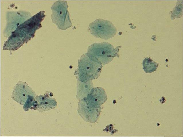
Likewise, a microscopic study of the vaginal discharge is done in order to detect the presence of key cells. These are epithelial cells in the vagina that are surrounded by bacteria..
Currently, the most accurate way to diagnose the disease is to perform a PCR test to genetically identify G. vaginalis.
G. vaginalis it is susceptible to various antibiotics such as ampicillin, carbenicillin, oxacillin, penicillin, and vancomycin. Strains have been observed to respond differently to tetracycline and gentaminycin, among others.
On the other hand, metrodinazole is quite effective. in vivo, but gives variable results in crops in vitro.
The most common treatments to treat the disease include the use of metronidazole or clindamycin. The application can be oral or vaginal creams.
In the case of oral application, metronidazole is usually used and the treatment lasts about seven days. When vaginal creams are applied, they can be based on metronidazole or clindamycin, which is applied for one to two weeks..
For pregnant patients with the disease, oral treatment is recommended, as it is considered safer and more effective.
These treatments can have some side effects such as nausea, stomach pains, cough and metallic taste in the mouth..
There are some alternative treatments, such as taking probiotics, that can help prevent relapse. Likewise, boric acid applications have shown some effectiveness.


Yet No Comments