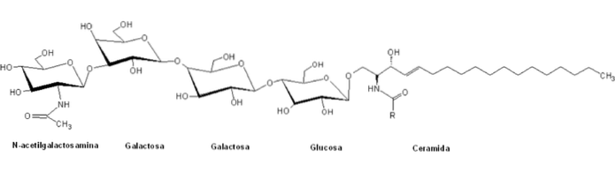
The globosides They are a type of sphingolipids belonging to the heterogeneous family of glycosphingolipids and are characterized by having in their structures a polar group composed of complex structured glycans linked to a ceramide skeleton by a B-glycosidic bond.
They are classified within the "globe" series of glycosphingolipids by the presence of a central structure of the general form Galα4Galβ4GlcβCer, and their nomenclature is generally based on the number and type of sugary residues of the polar heads.
Unlike other sphingolipids, globosides are normal constituents of the cell membranes of non-nervous systemic organs of many mammals. For example the kidneys, intestines, lungs, adrenal glands and red blood cells.
Like all membrane lipids, globosides have important structural functions in the formation and ordering of lipid bilayers..
However, and unlike their acidic or phosphorylated counterparts, the function of globosides is not related so much to the production of signaling molecules, but rather to their participation as part of glycoconjugates in the plasma membrane..
Article index
They share some structural and functional similarities with the other members of the group of glucosphingolipids: cerebrosides, gangliosides and sulfatides; including the composition of the main skeleton and the by-products of its metabolism.
However, globosides differ from acidic glycosphingolipids (such as gangliosides) with respect to the charge of their carbohydrate polar groups, since they are electrically neutral at physiological pH, which seems to have strong implications for their functions as part of the extracellular matrix..
These polar head groups normally have more than two sugar molecules, among which are commonly D-glucose, D-galactose and N-acetyl-D-galactosamine, and to a lesser extent fucose and N-acetylglucosamine.
As with other sphingolipids, globosides can be very diverse molecules, either taking into account the multiple combinations of fatty acids attached to the sphingosine skeleton or the possible variations of the oligosaccharide chains of the hydrophilic portion..
The pathway begins with the synthesis of ceramide in the endoplasmic reticulum (ER). The sphingosine skeleton is first formed by condensation of an L-serine and a palmitoyl-CoA.
Ceramide is subsequently generated by the action of ceramide synthase enzymes, which condense another molecule of fatty acid-CoA with the backbone of sphingosine at the carbon at position 2.
Still in the ER, the ceramides produced can be modified by the addition of a galactose residue to form galacto ceramides (GalCer), or they can instead be transported to the Golgi complex either by the action of ceramide transfer proteins (CERT ) or by means of vesicular transport.
In the Golgi complex, ceramides can be glycosylated to produce gluco ceramides (GlcCer).
GlcCer is produced on the cytosolic face of the early Golgi. It can then be transported to the luminal face of the complex and subsequently be glycosylated by specific glycosidase enzymes that generate more complex glycosphingolipids..
The common precursors of all glycosphingolipids are synthesized in the Golgi complex by the action of glycosyltransferases from GalCer or GlcCer.
These enzymes transfer specific carbohydrates from the appropriate nucleotide sugars: UDP-glucose, UDP-galactose, CMP-sialic acid, etc..
When GlcCer passes through the Golgi vesicular trafficking system it is galactosylated to produce lactosylceramide (LacCer). LacCer is the branch point from which the precursors of the other glycosphingolipids are synthesized, that is, the molecule to which more neutral polar sugar residues are subsequently added. These reactions are catalyzed by specific globoside synthases.
These lipids are mainly found in human tissues. Like many glycosphingolipids, globosides are enriched on the outside of the plasma membrane of many cells..
They are particularly important in human erythrocytes, where they represent the major type of glycolipid on the cell surface..
In addition, as noted above, they are part of the set of glycoconjugates of the plasma membranes of many non-nervous organs, mainly the kidneys..
The functions of globosides have not been fully elucidated to date, but it is known that some species increase cell proliferation and motility, in contrast to the inhibition of these events caused by some gangliosides..
A tetra-glycosylated globoside, Gb4 (GalNAcβ3Galα4Galβ4GlcβCer), functions in the site-sensitive recognition of structural disturbances of erythrocytes during cell adhesion processes..
Recent studies have determined the involvement of Gb4 in the activation of ERK proteins in carcinoma cell lines, which could mean its participation in tumor initiation. These proteins belong to the mitogen-activated protein kinase (MAPK) signaling cascade, consisting of the elements Raf, MEK, and ERK..
Their participation as receptors for some bacterial toxins of the Shiga family has been reported, specifically the globoside Gb3 (Galα4Galβ4GlcβCer), also known as CD77, expressed in immature B cells; also as receptors for HIV adhesion factor (gp120) and appear to have implications in certain types of cancer and other diseases.
There are numerous types of lipidosis in humans. Globosides and their metabolic pathways are related to two diseases in particular: Fabry disease and Sandhoff disease..
It refers to a sex-linked inherited systemic disorder, first seen in patients with multiple purple spots in the umbilical region. It affects organs such as the kidneys, heart, eyes, extremities, part of the gastrointestinal and nervous system.
It is the product of a metabolic defect in the enzyme ceramide trihexosidase, responsible for the hydrolysis of trihexosiceramide, an intermediate in the catabolism of globosides and gangliosides, which causes an accumulation of these glycolipids in the tissues.
This pathology was initially described as a variant of Tay-Sachs disease, related to the metabolism of gangliosides, but this also presents the accumulation of globosides in the viscera. It is an inherited disorder with autosomal recessive patterns that progressively destroys neurons and spinal cord.
It has to do with the absence of forms A and B of the enzyme β-N-acetyl hexosaminidase due to mutations in the gene HEXB. These enzymes are responsible for one of the degradation steps of some glycosphingolipids.


Yet No Comments