
GLUT1 is a transmembrane protein responsible for facilitating the passive transport of glucose across the plasma membrane, from the extracellular space to the interior of the cell.
In addition to glucose, it has been shown that it can also mobilize other six carbon sugars such as galactose, glucosamine and mannose. In turn, it allows the uptake and transport of vitamin C to the interior of cells unable to produce it..
Since all the molecules that are transported by GLUT1 are involved in the energy generation pathways in the cell, the expression of this transporter plays a very important metabolic role..
In fact, mutations that alter or abolish the expression of a functional GLUT1 result in the appearance of numerous diseases associated with slow neurological development and limited brain growth.
Article index
Glucose is the preferred carbon and energy source for most of the cells that make up the tree of life. Since it is not small enough and hydrophobic to cross cell membranes by itself, its transport into the cell requires the help of transporter proteins..
Two specific transporter-mediated transport mechanisms have been proposed for this sugar. One of them responds to a passive transport system (facilitated diffusion) and the second to an active transport system..
The first does not require energy to be carried out and occurs through a concentration gradient, that is, from a place of high glucose concentration to one where the concentration is lower..
Active glucose transport is carried out by transporters that obtain energy from sodium ion co-transport.
In contrast, the facilitated (passive) diffusion of glucose is carried out by a family of gate-like transporters called GLUT (for the acronym in English of “Glucose Transporters ”), family to which GLUT1 belongs. These bind glucose on the outside of the cell and transport it to the cytosol. At least 5 of them have been identified and their distribution appears to be different in different mammalian tissues..
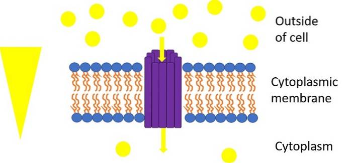
GLUT1 is a uniporter glucose transporter, that is, capable of carrying out the transport of glucose in only one direction, from the outside of the cell to the cytosol.
It belongs to the facilitated diffusion transporter (MSF) superfamily, which is widely distributed in many different organisms. It also participates in the transmembrane transport of a large number of small organic molecules..
Its peptide sequence of 492 amino acids is highly conserved in the different organisms where it has been identified, which is not difficult to believe given that the use of glucose to obtain energy constitutes the center of the metabolic tree of life..
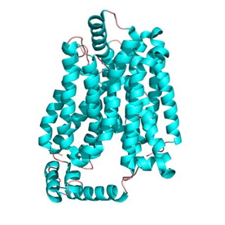
GLUT1 is an integral multipass membrane protein made up of 492 amino acid residues. This type of integral membrane proteins is characterized by crossing the lipid bilayer multiple times.
The three-dimensional chemical structure of proteins is generally determined through X-ray crystallography. The latter is a technique widely used by biochemists to reconstruct a structural model using pure crystals of the protein to be studied..
For highly conserved proteins such as GLUT1, determining the protein structure of a single organism may be sufficient. It is for this reason that researchers have so far determined the GLUT1 crystal structure of the mutant E3229..
As in all other members of the major facilitator superfamily (MSF), the structure of GLUT1 is represented by 12 transmembrane helices..
Additionally, in GLUT1 E3229, the amino and carboxyl terminal ends of the peptide are pseudo-symmetric and are oriented towards the cytosol. The arrangement of these ends generates a pocket or cavity that is open inside the cell and that constitutes the binding site for glucose..
Since glucose is generally transported from the outside to the inside of the cell, the finding that the binding site for this sugar is oriented towards the cytosol generates some confusion.
However, this confusion finds a solution in the results of biochemical investigations that suggest that a change occurs in the shape of the protein, allowing the glucose binding site to be exposed first on one side of the membrane and then on the other.
This does not mean that the protein rotates through the membrane, but rather that the binding of the sugar introduces the change in a way that, like a gate, exposes the glucose to the interior.
Since GLUT1 is a constitutive expression transporter, that is, it is always expressed in most mammalian cells, the functions it performs are vital for these cells. In fact, it is expressed in almost all tissues of the fetus precisely because during the development phases a high energy supply is needed to ensure growth..
However, its expression is decreased after birth in some tissues such as the liver, where the expression of other isoforms such as GLUT4 is now increased..
For erythrocytes it is of fundamental importance, since the latter depend exclusively on glucose for energy since they lack mitochondria. However, it is still responsible for the uptake of glucose to sustain respiration in the rest of cell types.
Since GLUT1 reaches a high concentration in the vascular endothelial cells of many organs and tissues, one of its functions is to carry glucose from the blood.
The transport of other hexoses such as mannose, galactose and glucosamine by GLUT1 does not question its direct relationship with energy metabolism, since ATP can be generated from all these hexoses.
In addition, the uptake and transport of vitamin C into cells unable to synthesize it has also been one of the functions reported for this ubiquitous receptor.


Yet No Comments