The hemianopia It is the loss of vision in one or both eyes, but only in half the visual field. Despite the peculiarity of this condition, it is not uncommon in medical practice, presenting in different degrees and with different characteristics, depending on the cause, severity and concomitants.
Etymologically speaking, the word has three components of Greek origin: hemi, which means "half"; an, which is a prefix related to "lack of" or "absence" and opsia, associated with "vision". The word would literally translate "lack of half vision" or "absence of half visual field".
Regardless of the origin of the disease, the common factor is optic nerve injury. Depending on which pathway is affected, on the same or contralateral side, one or another type of hemianopsia will appear. It should be remembered that all the fibers of the optic nerve are found in the chiasm, some of which cross and some of which do not..
The most common causes of this disorder are tumors in the central nervous system, head injuries, and cerebrovascular disease. Brain surgeries can also have as a complication some damage to the optic pathway that causes hemianopia. Some neurological and immunological diseases present with this picture.
Treatment for hemianopia will depend on the cause. Unfortunately, some cases have no cure and it is possible that they progress progressively, eventually leading to total loss of vision..
However, the majority of patients with hemianopia have the possibility of improvement if the origin of the disease is treated in time and in an adequate way..
Article index
As has been explained on other occasions and despite the possible confusion, it is important to clarify that hemianopia is a clinical sign, therefore it does not have its own symptoms. What it does have are particular characteristics that will depend on the disease that causes this partial loss of vision..
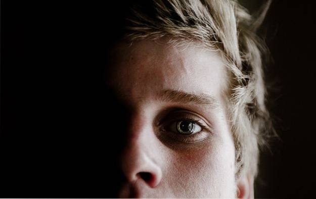
The fundamental characteristic of hemianopia is the decrease in visual acuity in the middle of the visual field. This decrease must be of such a degree that it is considered medically and legally blind. The most common is that it is the halves parallel to the nasal midline.
Patients with some type of hemianopia often have difficulty reading due to visual field compromise..
Walking is also impaired, frequently tripping over objects on the road or other people. In some cases there are hallucinations, flashing scotomas and pathological light effects.
As mentioned in the introduction, there are different causes of hemianopia, regardless of the visual half affected. The most important ones are mentioned below:
Many patients suffering from generalized polytrauma or head trauma have hemianopia among their symptoms..
These injuries can be acute, as in car accidents; or chronic, such as those suffered by many athletes in their practices (for example, boxers or American football players).
The time of appearance of symptoms in the latter will depend on the severity and frequency of the trauma. Hemianopia is usually permanent in these cases, since there is no well-defined solid lesion that can be tried to be extracted, but rather a cell, somatic or axonal damage..
Although the effect of the tumor is compressive or displacing, its behavior is similar to repeated trauma.
As the tumor grows it exerts pressure on certain brain structures, and if any of these has to do with the optic nerve, vision will be affected, almost always progressively.
The main difference with trauma is that these injuries are usually well defined in the brain anatomy. This does not mean that all of them can be operated on or resolved, but they offer a greater opportunity for improvement if the treatments, both medical and surgical, are started on time..
The previously called cerebrovascular accidents are a frequent cause of hemianopsia. If the area of the brain affected by the sudden cessation of blood supply - either by obstruction of the vessel that feeds it or by rupture of the same - fulfills visual tasks, it is possible that hemianopia or another vision alteration appears.
Although infrequent, cerebrovascular diseases typical of the vessels supplying the optic nerve can occur. Ischemic optic neuropathy can be anterior or posterior, depending on the segment of the nerve affected, with the anterior or frontal portion (also known as the head of the nerve) being the most commonly involved (90%).
Vascular migraines, a frequent pathology in the young population, can produce hemianopsia in its most severe presentation.
Many migraineurs report partial loss of vision during painful crises; this finding is transitory and disappears when the headache subsides.
This phenomenon seems to be related to the momentary interruption of blood flow to the optic nerve due to the vasoconstriction typical of migraine headaches..
Some authors also attribute the appearance of visual disorders during migraines to the inflammation of the brain tissue and nearby arteries that occurs in these cases.
Also known as retro-orbital neuritis, it is the inflammation of the optic nerve that causes loss of vision and pain when mobilizing the eye. In 90% of patients, only one eye is involved and it is always accompanied by changes in the pupillary response on the affected side..
Most cases are of idiopathic origin -that is, the cause is unknown-, although its relationship with other systemic pathologies has been proven.
Some of these diseases are multiple sclerosis, lupus erythematosus, Sjögren's syndrome, sarcoidosis, demyelinating neuropathies, and infections such as HIV / AIDS or mononucleosis..
The classification of hemianopia is simple and is based on two parameters: alteration of one or both eyes and compromised visual fields.
In this case, only one of the eyes is affected, regardless of which half is altered..
Both eyes are compromised, but not necessarily the same half on each side. In turn, this can be classified into:
It is the bilateral hemianopia in which both eyes have affected the same half of the visual field, either the right half or the left half.
In these cases, the optic nerve injury is located between the retina and the chiasm, so the loss of innervation is on the same side of the compromised visual field..
When the lesion is in the optic tracts, after the formation of the chiasm, the loss of vision affects the half of the visual field opposite the side of the lesion.
This type of hemianopia is called Contralateral Homonymous Hemianopia. Despite this, the same visual field is affected in both eyes.
As expected, in this type of hemianopia, the right side of the visual field is affected in one eye and the left side in the other. The half that is affected in one eye or the other, always different from each other, will depend on the location of the injury.
In this type of heteromeric hemianopia, the right half of the visual field of the left eye and the left half of the visual field of the right eye are affected..
To simplify the concept, it can be said that the two visual fields that look towards the nose are altered.
In this type of heteromeric hemianopia, the left half of the visual field of the left eye and the right half of the visual field of the right eye are altered. In short, vision is lost towards the ears on both sides.
As it is not a disease itself, but a symptom, the treatment will depend on the management of the causative pathology. For this same reason, the range of therapeutic alternatives is quite wide, including the following options:
Damage caused by certain tumors or acute trauma can be resolved through surgical procedures. The same occurs with the presence of intraparenchymal hematomas that compress the optic structures that cause hemianopia..
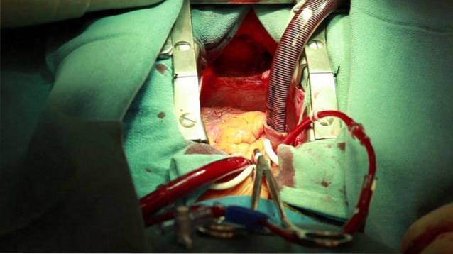
If the surgery is successful, the cure is usually immediate. However, some cases will require other complementary treatments that help to permanently regain vision..
If the cause of the hemianopsia is some medical pathology, such as those related to optic neuritis, the management is usually with medications or drugs.
Due to the same etiology of hemianopsia, steroids are the most frequently used drugs, since they help control the immune response and serve as anti-inflammatory drugs..
Certain visual exercises carried out with technological equipment, which stimulate the optic nerve endings, have been shown to be useful in the recovery of vision.
Other therapies, in which some auditory stimuli are also used together with visual stimuli in a harmonic way, improve the patient's conditions.
There are special glasses, created for each individual in particular, that have the ability to expand the patient's visual field. This is achieved through the insertion of some prisms in the lenses, which improve the patient's vision while wearing them..
Some patients receive specialized training to take advantage of healthy visual fields in both eyes. Patients incorporate these visual techniques into their daily life and report a significant improvement in the performance of their usual tasks.


Yet No Comments