
The hemolysin It is a small protein that causes pores in the cell membrane of erythrocytes and some other cells of the mammalian blood. It is generally synthesized and excreted by pathogenic bacteria.
This protein is one of the most common microbial toxins and the one that has been best studied. Sometimes it can cause hemolytic anemia, since the number of channels through which the cell interior exits can even cause cell lysis.
Generally, hemolysin is a typical toxin of the species of Streptococcus of the intestinal tract. Its function allows bacteria to break the epithelial barrier of the intestinal tract and thus move through the bloodstream to colonize other tissues..
The most common form that hemolysin is found in nature is in its α-hemolysin form. This protein is one of the most important virulence factors of most strains of Escherichia coli and some clostridia.
Most urinary tract infections are caused by strains of Escherichia coli producing α-hemolysin with hemolytic characteristics.
The production of hemolysin and bacteriocin in bacterial strains has been related to a competition mechanism against the other species and the production of both toxins seems to depend on the same genetic determinants in the genome of the bacteria..
Article index
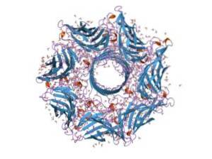
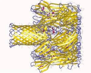
Hemolysin is made up of seven subunits and the gene that encodes it has seven promoters. These seven subunits insert themselves into the plasma membrane of the target cells and, when coming together, form an ion channel through which metabolites from the interior of the cell escape..
Hemolysin is an extracellular calcium (Ca + 2) -dependent cytotoxin that acts on the plasma membrane of bloodstream cells. The pores it creates in the membrane are also hydrophilic and cause water to enter the cell interior, which can lead to lysis..
Hemolysins are typical protein products of gram-negative bacteria and they all share two characteristics:
1- The presence of a very small peptide (nonapeptide) made up of repeats of the amino acids glycine and aspartic acid. The nonapeptides of hemolysin are located near the C-terminal portion of the primary structure of the protein.
2- All hemolysins are secreted by the bacteria to the extracellular medium through an ABC-type transporter (ATP-Binding Cassette).
Hemolysin production is usually detected in bacterial strains through growth on blood agar medium. In the test, a hemolytic halo is observed, a product of the breakdown of red blood cells near the bacteria colonies.
There are several different types of hemolysins, these are classified with a Greek letter at the beginning of their name. The most studied and common are α, β and γ hemolysins, all produced by the strain. Staphylococcus aureus.
The types of hemolysin are classified according to the range of cells they attack and according to their primary structure of the protein..
This protein is typical of strains of Staphylococcus aureus Y Escherichia coli; attacks neutrophils, red blood cells, lymphocytes, macrophages, adult and embryonic fibroblasts. Interacts with the polar heads of the plasma membrane lipids of these cells to internalize a hydrophobic tail of about 5 Ӑ inside the membrane.
Produced by Staphylococcus aureus To a lesser extent than α-hemolysin, β-hemolysin primarily attacks erythrocytes and enters the membrane exclusively through the sphingomyelin-rich domains of the cell membrane.
It has also been observed in Staphylococcus aureus. It has been classified as a hemolytic protein and a leukotoxin at the same time, since it affects polymorphonuclear cells of humans, monocytes, macrophages and rarely, even red blood cells.
This type of γ-hemolysin is one of the least characterized, therefore, much of its mechanism of action is unknown and it has not been investigated in vivo.
The mechanism of action that has been relatively clearly elucidated is that of α-hemolysin. However, since they are all hemolytic proteins, most of the processes are thought to be common to all hemolysins..
Scientists suggest that for bacteria to secrete hemolysin into the environment, they have to be in a nutrient-poor microenvironment, therefore, this would be a mechanism that triggers the cell to destroy the target cells and obtain their nutrients..
The mechanism has been described in three steps: cell membrane binding, insertion, and oligomerization..
Hemolysins have been found to be able to bind to neutrophil integrins, and in erythrocytes these proteins have been found to bind to glycosylated components such as glycoproteins, gangliosides, and cell membrane glycophorins..
Some authors suggest that the presence of receptors in the membrane is not essential for the binding of hemolysins to occur. In any case, the mechanism of cellular re-eating of the protein is not yet known with precision..
The interaction with the membrane occurs in two steps:
- Initial binding (reversible): when hemolysin binds to the calcium-binding domains of the membrane. This step occurs on the surface and is very susceptible to electrostatic discharge..
- Irreversible binding: joins the amino acid domains with the lipid components of the outer layer of the plasma membrane of the target cells, in order to form physical bonds between the hydrophobic compounds of the membrane.
Α-Hemolysin inserts residues 177 and 411 into the first lipid monolayer. In the extracellular environment, hemolysin is associated with calcium ions, which induce a structural arrangement in it and contribute to its activation..
This insertion consolidates the irreversible attachment to the cell membrane. Once the arrangement has occurred, hemolysin becomes an integral protein since, experimentally, it has been shown that the only way to extract it from the membrane is by using detergents such as Triton X-100.
When all the hemolysin has been inserted into the plasma membrane of the target cells, the oligomerization of the 7 subunits that make it up takes place, which ends in the formation of a protein pore, very dynamic but dependent on the lipid composition of the membrane..
It has been observed that the oligomerization process is favored by the microdomains or lipid rafts of the cell membrane. These regions may not favor the binding of the protein, but they do favor its oligomerization once inserted..
The more hemolysins that bind to the membrane, the more pores will form. Furthermore, hemolysins can oligomerize each other (adjacent ones) and form much larger channels..


Yet No Comments