The subarachnoid hemorrhage it is a spill of blood produced in the subarachnoid space. The latter is part of the cerebral meninges, and is the cavity through which the cerebrospinal fluid circulates. This liquid is responsible for protecting the brain from serious injury, as it serves as a cushion.
The subarachnoid space is located between the arachnoid layer and the dura mater, which are two of the three layers of the cerebral meninges. These are membranes that support, nourish, and protect the brain and spinal cord..
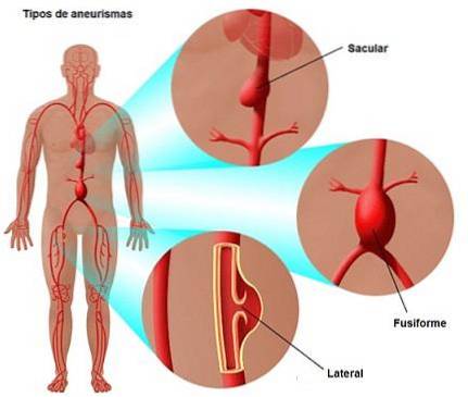
The first cause of a subarachnoid hemorrhage is a ruptured aneurysm (dilation of the walls of the arteries or veins). More rarely, it can be caused by an arteriovenous malformation.
Saccular aneurysms, that is, sac-like bulges in the wall of the arteries, are the most common. These correspond to 95% of aneurysms that rupture and that can cause subarachnoid hemorrhage..
Aneurysms generally originate in the arterial branches at the base of the brain. They can occur at or near the polygon of Willis (also called the cerebral arterial circle). The largest aneurysms are in the middle cerebral artery.
The areas most affected by aneurysms are: the junction of the carotid artery with the posterior communicating artery, the anterior communicating artery and the first bifurcation of the middle cerebral artery in the Silvio fissure.
Subarachnoid hemorrhage is a condition that can occur quickly and it is essential that the affected person receive immediate medical attention to ensure their survival. It usually occurs in people ages 40 to 60.
It has a mortality of up to 30% in the first month, even applying the most current treatments. Subarachnoid hemorrhage is a serious condition that can cause sequelae in 60% of patients. 40% of survivors are left in a state of dependency.
The incidence of subarachnoid hemorrhage is high in the United States, Finland, and Japan, while it is lowest in New Zealand and the Middle East..
The incidence is especially low in Indians and Africans from Rhodesia compared to Europeans, which can be explained by the lower rate of arteriosclerosis in these populations..
Article index
The rupture of an aneurysm is the main cause of subarachnoid hemorrhage, reaching 85% of non-traumatic causes. Other causes can be bleeding due to an arteriovenous malformation, bleeding disorders or the use of anticoagulants..
Subarachnoid hemorrhage can also be the cause of a traumatic injury due to a traffic accident or a fall..
There are different conditions that correlate with the formation of saccular aneurysms. For example: hypertension, arteriosclerosis (hardening of the walls of the arteries), vascular asymmetry in the circle of Willis, persistent headache, pregnancy-induced hypertension, long-term use of pain relievers, and history stroke relatives.
Although aneurysms are not congenital, although there is a certain degree of genetic disposition in their appearance, as occurs in other connective tissue diseases. Some families are known to have three or more members of the first or second degree who have had aneurysms..
Saccular aneurysms can develop from a lack of continuity of the smooth muscle of the middle layer at the bifurcations of the arteries. The artery wall protrudes through the muscle defect and the saccular formation or "bag" is generated..
The sacs have a thin wall of fibrous tissue. Clots and fibrin are deposited in these. It presents as a swollen balloon, and rupture occurs when there is intracranial pressure. This can appear for various reasons such as physical or emotional tension, lifting heavy objects, defecation or sex.
The risk of an aneurysm rupturing varies depending on its size. There is less risk in those that are less than 3 millimeters.
Subarachnoid hemorrhage can occur at any age, and some people are even born with aneurysms that can cause it. These patients must have continuous medical follow-up to prevent and control possible complications..
Women are more prone to subarachnoid hemorrhages than men. Other risk factors that increase the likelihood of suffering a subarachnoid hemorrhage include tobacco use, alcohol abuse, and high blood pressure..
Subarachnoid hemorrhage is a medical emergency that requires prompt attention. Healthcare personnel must be prepared to diagnose it and refer the patient to specialized centers for effective intervention.
- When a subarachnoid hemorrhage occurs there is a sharp increase in intracranial pressure. A sudden, severe headache occurs at first. Patients describe it as "the worst headache they have ever had" and that it can lead to loss of consciousness.
- Vomiting is also common, although nausea, phonophobia (sensitivity to noise) and photophobia (sensitivity to light) can occur separately..
- Seizures can occur by altering the electrical activity of the brain.
- On the other hand, there may be neck pain, numbness in the body, pain in one shoulder, confusion, irritability, and loss of alertness..
- On physical examination, stiffness in the neck may be found, although sometimes it only occurs hours after its appearance..
- Increased intracranial pressure can be transmitted to the area of cerebrospinal fluid that surrounds the optic nerves. This can lead to the rupture of veins in the retina, causing alterations in vision..
- During the first 2 or 3 days there may be an increase in body temperature, but it almost never rises above 39 degrees.
Other early neurological signs may also occur after subarachnoid hemorrhage and vary depending on the location of the aneurysm:
- Hemiparesis (weakness in only one half of the body), especially when there is an aneurysm in the medial cerebral artery.
- Paraparesis (slight difficulty in movement of the lower extremities): can occur when there is an aneurysm in the anterior communicating artery or a spinal arteriovenous malformation.
- Cerebellar ataxia (loss of muscle coordination due to involvement of the cerebellum): when there is a dissection of the vertebral artery.
- Third cranial nerve palsy (the oculomotor nerve, responsible for the eye muscles, is affected). It occurs when there is an aneurysm in the internal carotid artery, specifically at the beginning of the posterior communicating artery.
- Paralysis of the IX (glossopharyngeal nerve) and XIII cranial nerve (hypoglossal nerve responsible for coordinating the movements of the tongue): when there is a dissection of the vertebral artery.
Approximately 25-50% of patients die in the first rupture of the aneurysm, but a large part survive and improve in the following minutes. 4 or 9 days after the rupture, cerebral vasospasm (narrowing of the arteries) may occur.
Although it is one of the most common clinical pictures in neurology, errors in diagnosis are very frequent. It can be confused with migraine, meningitis, cerebral ischemia, hypertensive encephalopathy and emotional disorders.
Subarachnoid hemorrhage is often found on physical exam. The doctor may observe that patients have stiff neck and vision problems. Although to check it, you must perform other specific tests.
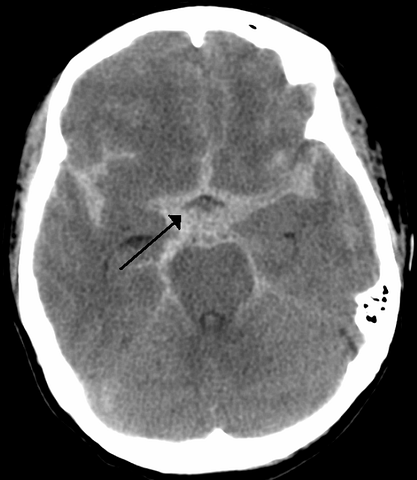
Subarachnoid hemorrhage is diagnosed by the presence of blood in the cerebrospinal fluid. This can be detected through a CT scan or lumbar puncture..
In 90% of cases, this sign can be seen if the scan is performed within the first 24 hours. If this test is negative, a lumbar puncture should be performed. This confirms subarachnoid hemorrhage if it is ruled out that a vessel has been injured when performing the puncture..
The CT scan helps locate the aneurysm and the area at risk for vasospasm. When there is a large amount of blood there is a greater risk.
After the CT scan, angiography of all four brain vessels should be performed. Usually this test does not show the cause of the bleeding, but if it is repeated in the following days, the aneurysm may be seen.
If it is not proven that it is an aneurysm, the most indicated is to perform an MRI to look for arteriovenous malformations in the brain, brainstem or spinal cord.
Electrocardiograms showing abnormalities or electrolyte studies of the blood should also be done. That is, an analysis to measure the level of minerals present in the blood or urine.
Also, to verify vasospasm, a transcranial Doppler examination (sound waves that allow images of the brain and cerebrospinal fluid) can be performed..
To determine that there is subarachnoid hemorrhage, a differential diagnosis is important. In other words, make sure that it is not being confused with other conditions such as epilepsy, metabolic encephalopathies, alcohol intoxication, tumors that lead to bleeding, meningitis, cervical osteoarthritis, cervical contractures ... among others..
Different scales are also used to measure the severity of subarachnoid hemorrhage according to its clinical manifestations. The most common with the Hunt and Hess scale, the Fisher scale and the scale of the World Federation of Neurological Surgeons.
Treatment is focused on excluding the aneurysm or vascular malformation from the circulation. It should be done immediately to prevent rebleeding.
This is achieved through surgery, slowing or decreasing the blood flow to the affected arterial vessel (embolization).
This can be done with catheter-guided balloons to open the blood vessels. Then "coils" are placed, which consist of small coils of soft metal. They are inserted into the aneurysm to block blood flow and prevent rupture.
Patients who are unable to undergo surgery should be treated until they can be operated on. This implies that they must be resting and with a central line (catheter).
People with significant neurological deficits should be admitted to the intensive care room. All measures to lower intracranial pressure should be used, including hyperventilation, use of mannitol (diuretic), and sedation.
The patient should be in a room with little light, isolated and with medications to prevent constipation, and pain relievers if necessary.
Seizures that generate new aneurysms may occur, therefore, the administration of anticonvulsants is necessary.
Vasospasms may also need to be treated. For this, drugs such as nimopidine or papaverine are used.
Another technique is transluminal dilation (dilation of an artery through a catheter with a balloon that inflates and deflates)..
Vasospasm can also be treated by inducing hypertension and hypervolemia. This should be done after operating the aneurysm, as it could cause a rebleeding.
Subarachnoid hemorrhage causes non-neurological complications that are the most frequent and that can cause death. These complications can be cardiac arrhythmias, lung edema, lung infections, kidney disorders and hyponatremia (low sodium level)..
On the other hand, neurological complications can be:
- Rebleeding: it occurs in 30% of cases in the first month. When there is rebleeding there is a mortality rate of 70%.
- Vasospasms: is the leading cause of mortality in subarachnoid hemorrhage.
- Hydrocephalus: the abnormal increase in the amount of cerebrospinal fluid in the brain. It occurs in 25% of cases.
All these damages can cause brain injuries due to the destruction of neurons.
Depending on the area of the brain affected, the person may suffer sequelae such as paralysis or weakness on one side of the body, balance problems, aphasias (problems producing or understanding speech), memory difficulties, impulse control problems, disinhibition, etc..
About 51% of people with subarachnoid hemorrhage die. While a third of the people who survive may become dependent.
Most deaths occur within 2 weeks, so after that period, the patient is most likely to survive. 10% of them before receiving medical attention and 25% within 24 hours of the bleeding. That is why it is important to see a doctor immediately..
The patient's level of consciousness upon admission, as well as the age and amount of blood from the hemorrhage are factors associated with a misdiagnosis..
The recovery period for subarachnoid hemorrhage is very long, and complications can arise if the patient is older or in poor health. In some cases, the treatment does not guarantee the improvement of the patient and some even die after this.
It should be emphasized that early care is essential. When a person presents the first symptoms of this condition, they should go urgently to a health center.


Yet No Comments