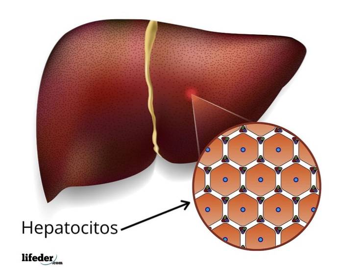
The hepatocytes They are the cells that make up liver tissue, that is, they are liver cells and participate in both the structure of the gland and its important exocrine and endocrine functions..
The liver is an essential organ for the human body. It is one of the largest glands and, in humans, it is located in the upper right quadrant of the abdominal cavity, just below the diaphragm. It weighs about 1.5 kg and is divided into 4 "portions" known as lobes..
The liver is highly irrigated by the circulatory system; In fact, about 12% of the blood volume of the human body is contained in this single organ, since part of its functions consist of filtering the blood, which is why it is also a target susceptible to the harmful effect of pathogens, fats, toxins and drugs.
The liver functions as an exocrine and endocrine gland:
Hepatocytes comprise most of the liver mass. They are relatively long-lived cells - renewed approximately every 5 months - and have a surprising capacity for proliferation and regeneration in the event of any damage..
Article index
Liver cells are responsible for the two main functions of this important gland:
These functions have a lot to do with the arrangement of hepatocytes in liver tissue, since they are both in contact with the blood capillaries of the liver (derived from the main veins), and with the bile canaliculi (where bile is excreted ).
In this context, we can say that hepatocytes function in:
Additionally, hepatocytes play an important role in another of the most important functions of the liver: the control of blood glucose levels..
To perform this function, these cells are responsible for internalizing glucose molecules derived from food and storing them in the form of glycogen, a polymer of glucose. Glycogen functions as an energy reserve and its catabolism releases glucose molecules into the blood when energy levels decrease.
Hepatocytes also function in the regulation of iron levels and in its storage in the form of ferritin; participate in the synthesis of cholesterol and various plasma proteins; act in the inactivation of hormones and fat-soluble drugs.
Another important function of these cells is the conversion of ammonium to urea and the conversion of amino acids and lipids into glucose through gluconeogenesis.,
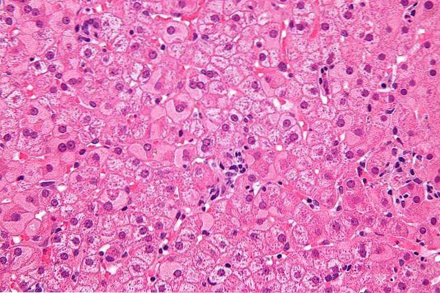
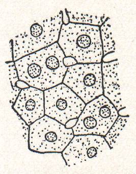
Hepatocytes are polyhedral cells, that is, they have several "sides" (usually 6) and these are usually flattened. It is through these "sides" that these cells come into contact with each other or with the hepatic sinusoids, which are the internal capillaries of the liver..
They are relatively large cells, since they can have diameters between 20 and 30 microns, with the average size of an animal cell being 20 μm.
Furthermore, they are polarized cells, which means that they have a "basal" and an "apical" region. The apical region is in contact with the bile canaliculi, which are the small ducts into which these cells excrete bile, while the basal region is in contact with the sinusoid spaces (capillaries).
The cytosol of hepatocytes usually has a granular appearance, since in addition to all the intracellular organelles, it contains hundreds of small stores of glycogen and lipids.
Hepatic cells have a central nucleus of variable size, although a small percentage of hepatocytes can be binucleated (with two nuclei).
Many of these cells have tetraploid nuclei (4n), that is, with twice the amount of DNA that other cells in the body have. These nuclei are usually larger than diploids (2n) and can have more than one nucleolar region.
Its rough endoplasmic reticulum is particularly abundant and participates in the fundamental tasks of the liver, such as the production of serum proteins (albumin, microglobulins, transferrin, ceruloplasmin and some components of lipoproteins).
The smooth endoplasmic reticulum, located between the rough endoplasmic reticulum and the Golgi complex, is also very abundant and its main functions are related to the presence of certain enzymes:
When observing liver cells under the microscope, it can be seen that many contain a system of well-defined saccules or cisterns, corresponding to the Golgi complex. In some it can be seen as a prominent membranous system that usually participates in:
Closely related to the membranes of the Golgi complex, lysosomes participate in the degradation of different intracellular materials, especially those that are potentially dangerous.
Hepatocytes also contain abundant peroxisomes - between 200 and 300 per cell - which are also involved in the detoxification of cells that have received toxic compounds from the blood..

Each liver cell can have between 100 and 800 mitochondria homogeneously distributed throughout the cytosol and exercising its main function: the synthesis of energy in the form of ATP molecules..
Hepatocytes comprise about 80% of all cells in the liver and, in this gland, these cells can be arranged well into sheets -plates- one cell thick or in strands of cells.
Generally, the cell plates connect to each other forming a spongy-looking tissue and are arranged radially around the central veins of the gland, while the cords do so around the sinusoid capillaries..
Virtually all liver cells are bathed by blood, since the liver is irrigated in such a way that the blood cell-plasma interface is extremely large, which allows the bidirectional flow of molecules between the intracellular and extracellular compartments..
It is important to note that hepatocytes differ from other epithelial cells in that they are not associated with a basement membrane. Instead, their basolateral membranes are surrounded by a low-density extracellular matrix secreted by the cells themselves, which facilitates the diffusion and exchange of molecules..

Yet No Comments