The pulmonary hilum It is the area of the lung where the structures that form the root of the viscera enter and exit. It is a hollow or depression area with a triangular shape that is located on the mediastinal aspect of both lungs, behind the cardiac impression, closer to the posterior pulmonary limit than the anterior.
The rib cage is separated from the lung by a two-layered membranous structure called the pleura. The hilum is where the parietal pleura (which covers the rib cage) connects with the visceral pleura (which covers the lung), forming the meeting point between the mediastinum and the pleural cavities.

It is important to differentiate the pulmonary hilum from the pulmonary pedicle. Although many authors speak of one or the other interchangeably as if it were the same structure, certain classic anatomy books and some purists of medicine continue to treat them as separate entities..
These anatomists refer to the hilum, not only of the lung but of any other organ, as the site of entry or exit of certain structures, but not the group of elements itself.
In this article, the hilum will be treated in its two nuances: as the entrance and exit door and as everything that enters or leaves the lung.
Article index
The components of the pulmonary hilum are those that form the pedicle or root of the lung itself. The root is wrapped in a thin, tubular-shaped layer of pleura that runs downward like a narrow fold, called the pulmonary ligament. This ligament serves as the link between the mediastinal and pulmonary parts of the pleura..
The pulmonary pedicle structures enter and exit through the hilum, allowing it to be connected to the heart and trachea.
This explains the support that the hilum provides to the pulmonary root, anchoring the lungs to the heart, trachea and other surrounding structures, providing firmness and protection to all the organs of the thorax..
Each hilum (and the respective root) is made up of:
- A main bronchus.
- A pulmonary artery.
- Two pulmonary veins.
- Bronchial arteries and veins.
- Pulmonary nerve plexuses (anterior and posterior).
- Lymphatic vessels.
- Bronchial lymphatic glands.
- Areolar tissue.
The right pulmonary root lies behind the superior vena cava and the right atrium, just below the azygos vein..
The upper lobe bronchus and the branch of the right pulmonary artery corresponding to the same lobe originate before entering the hilum, therefore they are seen above the level of the right main bronchus and the artery.
In the left hilum, the pulmonary artery occupies the upper part of the root, below which is the left main bronchus..
There are two pulmonary veins: one anterior and one posterior, with respect to the main bronchus. The rest of the structures closely resemble the right pulmonary hilum.
The main mission of the pulmonary hilum is to serve as an entry and exit port for the life-giving structures in the lung. In addition, thanks to the support of the pleura, it carries out support and protection functions for these structures, avoiding significant trauma, detachment and injuries or tears..
Clinically, the pulmonary hilum also provides information regarding the status and function of the lungs and other nearby structures..
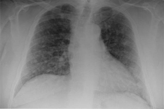
This task is accomplished thanks to the imaging studies that allow to observe or identify the pulmonary hila and their alterations or changes, such as X-rays, tomographies and resonances..
There are four basic reasons for a prominent or enlarged pulmonary hilum:
Cancer diseases such as lung cancer and lymphomas, as well as metastatic lesions from other primary tumors, can produce bulky masses in the hilar regions.
Lymphadenopathies also behave as masses that can appear in a widened hilum. Tuberculosis is the main infectious cause of pulmonary hilar lymphadenopathy, but not the only one; other viral, bacterial, and fungal infections often cause swelling of the hilar lymph nodes.
Some depot and autoimmune diseases are also responsible for causing widespread lymphadenopathy, including the lung area. Some drug reactions are even a relatively common cause of hilar lymphadenopathy..
Elevated pressure in the pulmonary veins can occur as a result of certain medical conditions. Heart failure and some types of valve disease -such as stenosis and mitral regurgitation- cause pulmonary venous hypertension, which is reflected as an increase in the size of the vessels and, therefore, hilar widening.
Other radiological evidences of pulmonary venous hypertension are interstitial edema due to plasma leakage into the lung parenchyma, ground glass appearance, peribronchial edema, and Kerley's B lines found in the lung bases and are signs of thickening suffered by the interlobular septa.
Elevated pressure in the pulmonary arteries can occur primarily or as a result of other systemic diseases. One of the most common causes is chronic obstructive pulmonary disease (COPD), which causes a significant increase in volume in the bilateral pulmonary hilum..
In newborns there is also a high risk of pulmonary hypertension due to respiratory maladjustment problems or congenital heart disease.
In them it is also possible to find signs of prominent pulmonary hilum in radiological studies together with other common findings, such as pruning of peripheral blood vessels.
Cyanogenic congenital heart diseases - in which there is a heart defect evident from birth that produces bluish or purplish discoloration of the skin and mucosa - can cause increased pulmonary blood flow and, consequently, widening of the pulmonary hilum.
As can be seen, there are a significant number of medical conditions that cause a prominent pulmonary hilum. After ruling out that it is an error in the radiological study, it is necessary to carry out the examinations and tests that the doctor considers necessary to properly diagnose and treat the cause..


Yet No Comments