
The hyperemia It is the redness and congestion of an anatomical region due to the accumulation of blood inside it. More than a disease, it is a symptomatic expression of some other clinical condition, and it is very important to determine the cause of hyperemia in order to decide if it is necessary to establish a particular treatment.
In some cases, the hyperemia is physiological, that means that the area is expected to be reddened due to a specific clinical or environmental circumstance. When this does not occur, that is, the tissue is not expected to be hyperemic, it is pathological hyperemia.
Hyperemia is a very common symptom that is usually associated with a local increase in temperature and sometimes pain, however these symptoms are not always associated.
Article index
Hyperemia is caused by vascular processes that cause the blood to be "dammed" in a certain area.
In this sense, arterial vasodilation may occur, which is responsible for a blood supply greater than normal to the hyperemic area. In these cases we speak of active hyperemia.
On the other hand, there may be the case of venous vasoconstriction which slows the outflow of blood from a certain area, therefore more red blood cells accumulate than normal and the area becomes red. When hyperemia is due to venous vasoconstriction it is known as passive hyperemia ".
There is a variant known as "reactive hyperemia" in which there is accumulation of blood in a certain area after a time of ischemia (absence of blood flow).
Although the conditions that can produce both active and passive hyperemia are multiple and varied, they all converge in a common mechanism: vasodilation (active hyperemia) or vasoconstriction (passive hyperemia).
The response on the blood vessels can be mediated by the autonomic nervous system (sympathetic: vasoconstrictor, parasympathetic: vasodilator), chemical mediators (vasoactive amines, prostaglandins) or a combination of both..
Although clinically they may be indistinguishable, there are various types of hyperemia according to their pathophysiology and within each group there are various causes.
A detailed explanation of each of them would take a whole volume of pathology, therefore emphasis will be placed on the most common types of hyperemia.
This is hyperemia that occurs under normal conditions. It is not associated with any disease and has no negative impact on the person who presents it.
Physiological hyperemia is a normal reaction to certain internal or external stimuli which result in vasodilation of the arterial capillaries.
One of the situations where physiological hyperemia is seen more frequently is in very hot environments. In such circumstances the body needs to dissipate heat to maintain its stable temperature and for this the capillaries of the skin expand allowing heat to be released as if it were a radiator.
When this happens, the skin becomes red, spontaneously returning to its normal condition as soon as the ambient temperature drops..
Another similar situation is during physical activity. In this case the mechanism is exactly the same, only that the heat instead of coming from the outside does it from the inside of the body, secondary to the muscular work. Once again the cutaneous capillaries dilate making the skin (especially the thinner skin of the face) look red.
Finally, in response to certain substances such as adrenaline (secreted by the body when faced with certain stimuli and emotions), the capillaries of the skin dilate causing it to turn reddish; a phenomenon known as "blush" or "blush".
In all these cases, the hyperemia is normal, harmless and temporary, the skin taking its normal color once the stimulus that produced the hyperemia ceases..
It is that type of hyperemia that constitutes a symptom of disease or pathological condition. Pathological hyperemia can be divided into active, passive and reactive..
Any clinical condition during which vasodilation of the arterial capillaries occurs will be associated with active hyperemia.
One of the typical and most frequent examples is fever. During febrile episodes, body temperature increases, as does the heart rate (hyperdynamic state of the blood), with vasodilation of arterial capillaries being associated as a compensatory mechanism for temperature. That is why people with a fever look flushed.
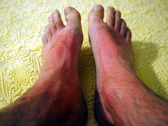
Something similar happens with first degree sunburn. Thermal injury increases local temperature causing arterial capillaries to dilate and give the skin a reddish hue. Chemical mediators such as interleukins, secreted in response to cell damage by solar radiation, are also associated at this point..
Interleukins have vasodilatory properties so in the presence of a sunburn or any other type of injury (trauma, infection, inflammation of any kind) they induce arteriolar vasodilation and therefore hyperemia.
From the above, it can be deduced that any situation where tissue damage occurs may be associated with active hyperemia, with frequent associated symptoms being swelling (due to increased capillary permeability in the area) and local increase in temperature..
Passive hyperemia occurs when, due to some condition, the venous capillaries contract, slowing down the drainage of blood from a given anatomical area..
A classic example is when a person spends a lot of time leaning on his arm or leg in a certain position. After a while the point of support becomes red. This occurs simply because the pressure when resting on that area occludes the venous capillaries so that the blood can enter but not leave, therefore that part of the anatomy turns red.
Although all cases of hyperemia in the skin have been described so far, from the anatomopathological point of view this condition can also occur in the internal organs.
In these cases, passive hyperemia is called "congestive hyperemia" which is nothing more than the accumulation of blood in a viscera due to the inability to adequately drain the blood..
This occurs frequently in congestive heart failure where the heart is not able to mobilize all the blood in the body efficiently, so it remains dammed in the peripheral organs, especially the liver and spleen..
It is the most common type of hyperemia in patients with arterial disease. Reactive hyperemia occurs when, after a more or less prolonged period of ischemia (insufficient blood supply to a limb or organ), normal blood flow is restored.
During ischemia, the arterial capillaries dilate as much as they can in order to supply as many red blood cells (and therefore oxygen) to the tissues they supply. As ischemia is maintained over time, more and more capillaries dilate in an attempt to keep the oxygen supply constant, however due to flow obstruction (which produces ischemia) the limb remains pale.
Now, once normal blood flow is restored, the capillaries do not contract ipso facto, in fact it takes a few hours, even days (depending on the previous ischemia time) for the arterial capillary bed to return to normal..
However, since the blood supply to the area increased, now the skin looks reddened since through the dilated capillaries where almost no blood circulated before, now it does so in huge quantities.
Since it is a symptom, hyperemia itself does not present complications, although the same cannot be said of the conditions it produces..
Thus, the complications of hyperemia are those of the condition that produces it; for example, in active hyperemia secondary to sunburn, the complications of hyperemia will be those associated with said type of burn.
On the other hand, if the hyperemia is due to fever or a skin infection (cellulitis), complications can be expected from either the fever or the infection..
The same goes for passive hyperemia. When a person presents passive hyperemia over a support area due to reduced mobility, it is expected that the hyperemia will sooner or later be associated with an eschar (pressure ulcer), so that in this case the complication is that derived from the mobility limitation.
This dissertation can be done one by one with all the causes of hyperemia so that as a corollary it is enough to remember, as previously stated, that the complications of hyperemia are those associated with the condition that causes it..
As with complications, there is no specific treatment for hyperemia, in this sense the definitive treatment should be aimed at improving, alleviating or eliminating the initial condition that caused the hyperemia..
However, there are general measures that can help alleviate the symptoms in most cases, in this sense the application of local cold by means of ice packs, ice pack or cold lotions is a common, effective and economical solution..
On the other hand, in cases of hyperemia secondary to histamine release (as in allergic reactions or stings of some insects), the administration of H1 blockers is of great help..
In general, it can be concluded that the treatment of hyperemia is based on three pillars:
- Eliminate exposure to the causative agent (if possible).
- Control as much as possible the underlying condition that produced the hyperemia.
- Symptomatic treatment by administering general palliative measures.


Yet No Comments