
The hypotrophy It can be defined as a delay in the development of a tissue or an organ without altering its structure. It can occur, in some cases, as an involutive process due to reducing use, work, nervous, hormonal, blood stimulation or due to aging..
It can also be defined as the degeneration in the functioning of an organ due to the reduction in the size of its cells or the loss of the number of cells. Some authors consider hypotrophy as a synonym for atrophy, while others consider atrophy as the maximum degree of hypotrophy..
Knowledge related to the functional and structural reactions of cells and tissues to agents capable of causing injuries, including genetic defects, is the key to understanding pathological processes..
Diseases are currently defined and interpreted in molecular terms and not just as a general description of structural alterations. Cellular and biological tissue alterations can be the result of adaptations, injuries, neoplasms, age or death.
Article index
Adaptations can occur as a normal or physiological response, or as a consequence of an adverse or pathological situation. The most significant adaptive cell or tissue changes include:
-hypotrophy or atrophy, which is a decrease in the size of cells.
-hypertrophy or increase in cell size.
-hyperplasia or increase in the number of cells.
-metaplasia, which is the reversible replacement of a mature cell by another immature type.
-dysplasia, which is a disorderly growth and is considered more than a cellular adaptation, an atypical hyperplasia.
Hypotrophy or atrophy is, therefore, a process of cellular adaptation and in this text the two terms will be considered as synonyms.
Atrophy or hypotrophy consists of a decrease or contraction of cell size. If the process occurs in a significant number of cells in an organ, the entire organ shrinks and becomes "hypotrophic" or "atrophic," reducing its function..
Although this process can affect any organ, it is much more frequent in the skeletal muscles and the heart and, secondarily, in the sexual organs and the brain..
Hypotrophy can be classified as physiological or pathological. Physiological can occur early in development. For example, the thymus atrophies early in childhood. Pathological occurs as a result of decreased workload, use, pressure, blood supply, nutrition, and hormonal or nervous stimulation..
People who are immobilized in bed suffer from disuse atrophy, age causes atrophy of neurons and endocrine organs, etc. In either case, whether physiological or not, hypotrophic cells exhibit the same basic changes.
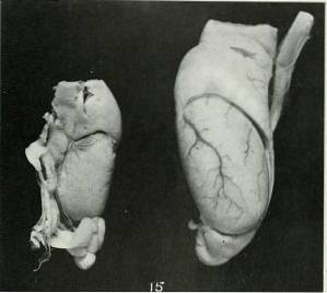
In hypotrophy or renal atrophy, the affected kidney is smaller than the normal kidney. This implies kidney dysfunction, that is, kidney disease that can have different causes. Among the most frequent causes are vascular problems and those related to the urinary system.
One of the most important vascular causes is renal ischemia, when the kidneys receive an insufficient amount of blood. The reduction in flow may be due to the presence of a clot that obstructs the lumen of the artery, it may be a problem of the arterial wall or external compressions due to cysts or tumors..
In the case of the urinary system, a significant obstruction in the elimination of urine can occur, which causes a retrograde accumulation to the site of obstruction and an increase in pressure with decreased renal function. The most common cause is stones.
Whatever the cause of hypotrophy, it must be corrected quickly before kidney damage is irreversible. Generally, these pathologies are accompanied by florid symptoms similar to those that occur in urinary tract infections..
Other times they are asymptomatic and there is no significant alteration in the final function, since the healthy kidney can compensate for the failure. In these cases, irreversible damage is very likely to occur and, as a consequence, the loss of the affected kidney..
In muscle hypotrophy, if atrophic muscle cells are compared with normal muscle cells, the former contain less sarcoplasmic reticulum, fewer mitochondria, and myofilament content is reduced.
If the atrophy was caused by loss of nerve connections, oxygen consumption and amino acid uptake are rapidly reduced.
This process appears to be accompanied by a reduction in protein synthesis or an increase in protein catabolism in affected cells, or both. The degradation pathway includes ubiquitin binding and involvement of proteasomes or proteolytic cytoplasmic complexes..
When the muscle remains shortened to a length less than its normal length and this occurs continuously, the sarcomeres at the ends of the muscle fibers rapidly disappear. This is part of a muscle remodeling mechanism, which aims to establish the optimal length for contraction..
Testicular hypotrophy can have a genetic origin, it can occur as a consequence of aging, or it can have a frank pathological cause. It is characterized by a decrease in testicular size and can be unilateral or bilateral.
The sperm count decreases and there is a decrease in the size and number of Leydig cells (producing testosterone) and germ cells (producing sperm).
Klinefelter syndrome, which is a syndrome of genetic origin that only affects males, is accompanied by testicular atrophy, sterility, hyalinization of the seminiferous tubes and gynecomastia.
The decrease in testosterone levels that occurs in old age leads to a decrease in the size of the testicles and a reduction in sexual drive.
Among the most frequent pathological causes are varicocele, testicular cancer, orchitis, chronic and excessive alcohol consumption, the use of hormones such as anabolic steroids, the administration of estrogens and testicular torsion, among others..
Uterine hypotrophy is a uterine feature of the post-menopausal period. The uterus is reducing in size, shrinking and, around the age of 65, it can be seen frankly atrophic, concomitantly atrophy of the ovaries and vagina occurs.
The changes in the uterus and vagina are due to the decrease in estrogen levels that occurs in female menopause. The use of medications that block or inhibit estrogen functions can lead to uterine and vaginal atrophy.
Brain hypotrophy is a common condition in many pathologies that affect brain tissue. It consists of a decrease in the size of the cells that leads to a decrease or reduction in the size of the organ. In the case of brain tissue, this implies the loss of neurons and / or their connections..
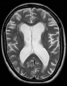
Symptoms include changes in mood, personality, and behavior. It can present as dementia, spatial and / or temporal disorientation, memory loss, learning problems, difficulty with abstract thoughts, problems speaking, reading and understanding, among others..


Yet No Comments