The steroid hormones They are substances made by glands of internal secretion and that are dumped directly into the circulatory stream, which leads them to the tissues where they exert their physiological effects. Its generic name derives from the fact that it has a steroidal nucleus in its basic structure..
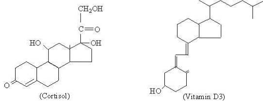
Cholesterol is the precursor substance from which all steroid hormones are synthesized, which are grouped into progestagens (for example progesterone), estrogens (estrone), androgens (testosterone), glucocorticoids (cortisol), mineralocorticoids (aldosterone) and vitamin D.
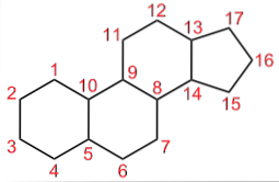
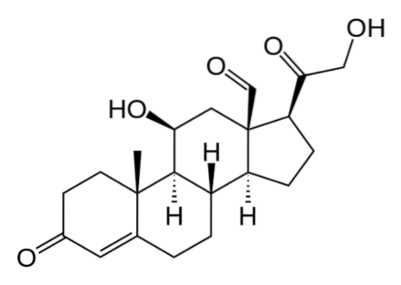
Although the different steroid hormones present molecular differences between them, which are what give them their different functional properties, it can be said that they have a basic structure that is common to them and that is represented by the cyclopentaneperhydrophenanthrene of 17 carbon atoms.
Article index
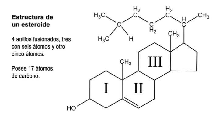
Steroids are organic compounds of a very diverse nature that have in common what could be considered as a parent nucleus consisting of the fusion of three rings of six carbon atoms (cyclohexanes) and one of five carbon atoms (cyclopentane).
This structure is also known as "cyclopentaneperhydrophenanthrene". Since the rings are mutually linked, the total number of carbon atoms that make it up is 17; However, most natural steroids have methyl groups at carbons 13 and 10, which represent carbons 18 and 19, respectively..
Many of the natural steroidal compounds also have one or more groups with alcoholic function in the ring structure and are therefore called sterols. Among them is cholesterol, which has an alcohol function at carbon 3 and a hydrocarbon side chain of 8 carbon atoms attached to carbon 17; atoms that are numbered from 20 to 27.
In addition to these 17 carbons, steroid hormones can have 1, 2 or 4 more of these atoms in their structure, for which three types of steroids are recognized, namely: C21, C19 and C18.
The C21s, like progesterone and adrenal corticosteroids (glucocorticoids and mineralocorticoids), are derived from “pregnane”. It has 21 carbon atoms because to the 17 of the basic ring are added the two of the methyl groups of carbons 13 and 10, and two carbons of the side chain attached to C17 that originally, in cholesterol, was 8 carbons.
The C19s correspond to sex hormones with androgenic activity and are derived from “androstane” (19 carbon atoms), which is the structure that remains when pregnane loses the two carbons of the C17 side chain, which is replaced by a hydroxyl or a ketone group.
C18 steroids are female hormones or estrogens that are synthesized mainly in the female gonads and whose outstanding characteristic, with respect to the other two types of steroids, is the absence of the methyl present in the latter attached to the carbon in position 10.
During the synthesis from cholesterol, enzymatic modifications are produced that alter the number of carbons and promote dehydrogenations and hydroxylations of specific carbons of the structure..
The cells that produce steroid hormones are located mainly in the cortex of the adrenal glands, where glucocorticoids such as cortisol, mineralocorticoids such as aldosterone, and male sex hormones such as dehydroepiandrosterone and androstenedione are produced..
Male sex gonads are responsible for the production of androgens, including the aforementioned hormones and testosterone, while the maturing ovarian follicles produce progesterone and estrogens..
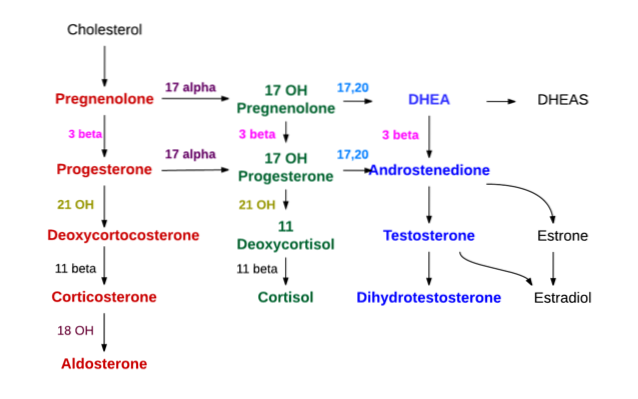
The synthesis of all steroid hormones starts from cholesterol. This molecule can be synthesized by cells that produce steroid hormones, but for the most part it is obtained by these cells from low-density lipoproteins (LDL) present in circulating plasma..
In the adrenal cortex, three layers are distinguished, known from the outside in as the glomerular, fascicular and reticular zones, respectively..
In the glomerular, mineralocorticoids (aldosterone) are mainly synthesized, in the fascicular glucocorticoids such as corticosterone and cortisol, and in the reticular androgens such as dehydroepiandrosterone and androstenedione.
The first step in the synthesis occurs in the mitochondria and consists of the action of an enzyme called cholesterol desmolase, belonging to the cytochrome P450 superfamily and also known as "P450scc" or "CYP11A1", which promotes the elimination of 6 of the carbon atoms of the side chain attached to C17.
With the action of desmolase, cholesterol (of 27 carbon atoms) is converted into pregnenolone, which is a compound with 21 carbon atoms and represents the first of the C21 type steroids.
Pregnenolone moves to the smooth endoplasmic reticulum, where by action of the enzyme 3β-hydroxysteroid dehydrogenase undergoes dehydrogenation in the hydroxyl of the alcohol group of carbon 3, and becomes progesterone.
Due to the action of 21β-hydroxylase, also called “P450C21” or “CYP21A2”, progesterone is hydroxylated at carbon 21 and is transformed into 11-deoxycorticosterone, which returns to the mitochondria, and to which the enzyme 11β-hydroxylase (“ P450C11 ”or“ CYP11B1 ”) converts to corticosterone.
Another line of synthesis in the fascicular zone and that ends not in corticosterone, but in cortisol, occurs when pregnenolone or progesterone are hydroxylated in position 17 by 17α-hydroxylase ("P450C17" or "CYP17") and converted into 17-hydroxypregnolone or 17-hydroxyprogesterone.
The same enzyme already mentioned, 3β-hydroxysteroid dehydrogenase, which converts pregnenolone to progesterone, also converts 17-hydroxypregnolone to 17-hydroxyprogesterone.
The latter is carried successively, by the last two enzymes of the pathway that produces corticosterone (21β-hydroxylase and 11β-hydroxylase) to deoxycortisol and cortisol, respectively..
The main glucocorticoids produced in the zona fascicular of the adrenal cortex are corticosterone and cortisol. Both substances, but especially cortisol, display a broad spectrum of actions that affect metabolism, blood, defense and wound healing responses, bone mineralization, the digestive tract, the circulatory system, and the lungs..
Regarding metabolism, cortisol stimulates lipolysis and the release of fatty acids that can be used in the liver for the formation of ketone bodies and low-density proteins (LDL); decreases glucose uptake and lipogenesis in adipose tissue and glucose uptake and utilization in muscle.
It also promotes protein catabolism in the periphery: in connective tissue, muscle and bone matrix, thereby releasing amino acids that can be used in the liver for the synthesis of plasma proteins and for gluconeogenesis. Additionally stimulates intestinal glucose absorption by increasing the production of SGLT1 transporters.
Accelerated intestinal glucose absorption, increased hepatic production and decreased utilization of this carbohydrate in muscle and adipose tissue favors an elevation in plasma glucose levels..
Regarding the blood, cortisol favors the clotting process, stimulates the formation of neutrophil granulocytes and inhibits that of eosinophils, basophils, monocytes and T lymphocytes. It also inhibits the release of inflammatory mediators such as prostaglandins, interleukins, lymphokines, histamine and serotonin.
In general, it can be said that glucocorticoids interfere with the immune response, for which reason they can be used therapeutically in those cases in which this response is exaggerated or inappropriate, such as in the case of autoimmune diseases or in organ transplants to reduce rejection.
Androgen synthesis at the level of the adrenal cortex occurs mainly at the level of the reticular zone and from 17-hydroxypregnolone and 17-hydroxyprogesterone.
The same 17α-hydroxylase enzyme, which produces the two substances just mentioned, also has 17,20 lyase activity, which removes the two carbons of the C17 side chain and replaces them with a keto group (= O).
This last action reduces the carbon number by two and produces C19 type steroids. If the action is on 17-hydroxypregnolone, the result is dehydroepiandrosterone; if, on the contrary, the affected substance is hydroxyprogesterone, then the product will be androstenedione.
Both compounds are part of the so-called 17-ketosteroids, as they have a ketone group at carbon 17.
3β-hydroxysteroid dehydrogenase also converts dehydroepiandrosterone to androstenedione, but the most common is that the former is converted to dehydroepiandrosterone sulfate by a sulphokinase, present almost exclusively in the reticular zone.
The zona glomerularis lacks the 17α-hydroxylase enzyme and cannot synthesize the 17-hydroxysteroid precursors of cortisol and sex hormones. It also does not have 11β-hydroxylase, but it does have an enzyme called aldosterone synthetase that can sequentially produce corticosterone, 18-hydroxycorticosterone, and the mineralocorticoid aldosterone..
The most important mineralocorticoid is aldosterone synthesized in the zona glomerularis of the adrenal cortex, but glucocorticoids also display mineralocorticoid activity.
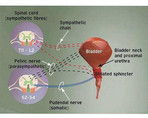
The mineralocorticoid activity of aldosterone develops at the level of the tubular epithelium of the distal nephron, where it promotes the reabsorption of sodium (Na +) and the secretion of potassium (K +), thus contributing to the conservation of the levels of these ions in the body fluids.
Testicular androgen synthesis occurs at the level of Leydig cells. Testosterone is the main androgen hormone produced in the testes. Its synthesis involves the initial production of androstenedione as previously described for the synthesis of androgens at the level of the adrenal cortex..
Androstenedione is converted into testosterone by the action of the enzyme 17β-hydroxysteroid dehydrogenase, which replaces the ketone group of carbon 17 with a hydroxyl group (OH).
In some tissues that serve as a target for testosterone, it is reduced by a 5α-reductase to dihydrotestosterone, with greater androgenic power.
This synthesis occurs cyclically accompanying the changes that occur during the female sexual cycle. The synthesis occurs in the follicle that during each cycle matures to release an ovum and then produce the corresponding corpus luteum..
Estrogens are synthesized in the granular cells of the mature follicle. The mature follicle has cells in its theca that produce androgens such as androstenedione and testosterone..
These hormones diffuse into neighboring granulosa cells, which possess the aromatase enzyme that converts them to estrone (E1) and 17β-estradiol (E2). From both, estriol is synthesized.
Androgens and estrogens have as their main function the development of male and female sexual characteristics respectively. Androgens have anabolic effects promoting the synthesis of structural proteins, while estrogens favor the ossification process.
The estrogens and progesterone released during the female sexual cycle are intended to prepare the woman's body for an eventual pregnancy as a result of the fertilization of the mature egg released during ovulation..
If you need to refresh your memory about the mechanism of action of hormones, it is recommended to watch the following video before continuing reading.
The mechanism of action of steroid hormones is quite similar in all of them. In the case of lipophilic compounds, they dissolve without difficulty in the lipid membrane and penetrate the cytoplasm of their target cells, which have specific cytoplasmic receptors for the hormone to which they must respond..
Once the hormone-receptor complex is formed, it crosses the nuclear membrane and binds in the genome, in the manner of a transcription factor, with a hormone response element (HRE) or primary response gene, which in turn It can sometimes regulate other genes called secondary response.
The end result is the promotion of transcription and the synthesis of messenger RNAs that are translated in the ribosomes of the rough endoplasmic reticulum that end up synthesizing the proteins induced by the hormone..
The action of aldosterone is mainly exerted at the level of the final portion of the distal tube and in the collecting ducts, where the hormone promotes Na + reabsorption and K secretion+.
In the luminal membrane of the main tubular cells of this region, there are epithelial Na + channels and K + channels of the "ROMK" type. Renal Outer Medullary potassium Channel).
The basolateral membrane has Na + / K + ATPases pumps that continuously draw Na + from the cell into the basolateral interstitial space and introduce K + into the cell. This activity keeps the intracellular concentration of Na + very low and favors the creation of a concentration gradient for this ion between the lumen of the tubule and the cell..
Said gradient allows Na + to move towards the cell through the epithelial canal, and since Na + passes alone, for each ion that moves, an uncompensated negative charge remains that causes the lumen of the tubule to become negative with respect to the interstitium. That is, a transepithelial potential difference is created with the negative light.
This negativity of light favors the exit of K + which, due to its higher concentration in the cell, and the negativity of light is secreted towards the lumen of the tubule to be finally excreted. It is this Na + reabsorption and K + secretion activity that is regulated by the action of aldosterone..
Aldosterone present in the blood and released from the zona glomerularis in response to the action of angiotensin II, or to hyperkalemia, penetrates into the main cells and binds with its intracytoplasmic receptor.
This complex reaches the nucleus and promotes the transcription of genes whose expression will end up increasing the synthesis and activity of Na + / K + pumps, epithelial Na + channels and ROMK K + channels, as well as other proteins. Response that will have the overall effect of the retention of Na + in the body and the increase in urinary K excretion+.
Yet No Comments