
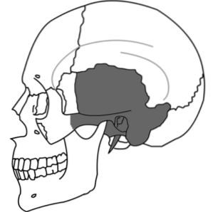
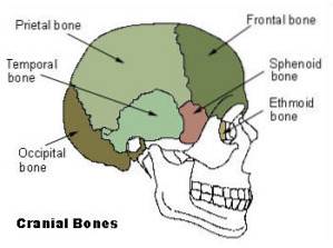
The temporal bone It is a bone structure that is part of the skeleton of the cranial vault. It is an even bone that is in a lateral-medial position and extends to the lower part of the skull.
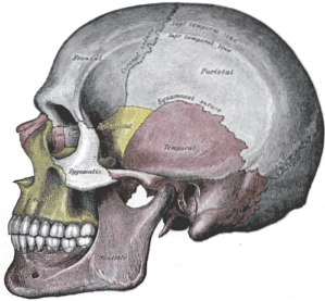
It is related to the parietal, occipital and sphenoid bone, with which it forms joints and cranial lines called sutures. During its development in the fetus, the bone consists of three separate portions that later join together to form a single, solid structure in the newborn.
The temporal bone is responsible for protecting important vascular and neurological structures, including the internal carotid artery, jugular vein, and internal organs of hearing..
Although the temporal bone is a thick and strong bone and requires significant trauma to fracture, when these injuries occur they have a high degree of complications and can even lead to death..
When a polytraumatized patient presents with vertigo, bleeding through the ears or refers to difficulty hearing, a lesion of the temporal bone should be suspected and should begin to be evaluated by imaging studies such as magnetic resonance imaging (MRI) and computerized axial tomography (TAC).
Article index
The temporal bone is a paired bony structure found in the lateral part of the skull. Be part of neurocranium, which are those bones that are found in the upper part of the cranial vault.
It is divided into four parts for your better anatomical understanding. In the embryo, these parts are totally independent but fuse before birth.
These parts are called: squamous portion, petrous portion, mastoid portion and tympanic portion..

Although the temporalis is part of the neurocranium, it extends to the lower part of this structure and, together with the ethmoid, sphenoid, occipital bones and the basal portion of the frontal bone, it forms the base of the skull..
The amount of structures found at its level make it an important protective shield against external trauma. It is a strong bone and its fracture is difficult.
Its main function is to protect the brain. Together with the neighboring bone structures, it is responsible for protecting the important neurological and vascular elements found within it..
It is a very important bone structure as it contains the organs of hearing, balance and the mandibular articular surfaces..
Its injury represents a danger to the quality of life of the patient, and can even cause death, since in addition to containing the organs of hearing and balance, it contains most of the cranial nerves.
Nerves or cranial nerves are neurological structures that emerge directly from the brain and have important motor and sensory functions throughout the body, including breathing..
The future skull begins its formation around 4ta gestation week. At that time, the bone-forming cells begin the development of the structures corresponding to the cranial vault..
The bony nuclei that will form the temporal bone begin their development on the 6thta week. The cartilaginous part or chondrocranium, which gives rise to the bony structures at the base of the skull.
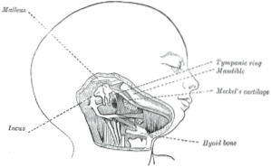
The calls ear capsules are the structures that will become the petrous and mastoid portion of the temporal bone.
Ossification of the temporal bone, or bone development from cartilage, begins at 16ta week, with the formation of the so-called temporal tympathetic rings. The petrous portion concludes its ossification in the 19na week.
It is important to emphasize that complete ossification of the chondrocranium does not occur until the vascular and neurological structures are fully formed, since the base of the skull gives way to all these elements. Then, once those structures are formed, the bone is molded around them.
At the time of birth, the three portions of the temporal bone have already been unified to form a single bone.
However, the rest of the skull bones are barely united by a fibrous, strong and elastic tissue, without having fused. These unions are called sutures.
The function of the sutures is to allow the skull to pass through the birth canal without posing a risk to the pregnancy product. In addition, after birth, it allows the correct development of the brain, finally merging towards the second year of life..
The ear is fully formed in the newborn and quickly expels the fetal fluid that has filled the spaces that make it up, replacing it with air..
The temporal bone is a complex structure that is divided into four parts and two protrusions. This division allows its better understanding for its anatomical study.
The names and general division of the different portions are due to the embryological development of the temporal bone, which begins as separate cartilaginous structures that develop individually to finally fuse into one solid bone..
The portions of the storm are as follows:
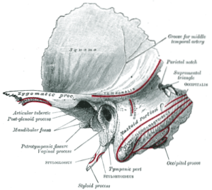
It is the largest part of the bone. It is also known as temporary scale or temporary shell. It is shaped like a convex plate and is located on the top and side of the skull. It has an external and an internal face.
On the surface of the external face A groove is observed that allows the passage of the posterior deep temporal artery. It also has a depression, located in the lower part, called mandibular fossa. This is where the temporal bone articulates with the jaw..
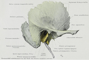
The internal face is concave, presents depressions that are formed by the cerebral convolutions and also has vascular furrows through which branches of the middle meningeal artery pass..
The zygomatic process is one of the protrusions that project from the lower part of the squamous portion of the temporal bone and articulates with the zygomatic bone, which is part of the face..
It is located posterior to the squamous portion. At its posterior edge, it is in contact with the occipital bone and a protrusion called the mastoid process is evident in that area.
This protrusion is evaluable on physical examination, it can be felt behind the ear. It is the site of insertion of various muscles, such as the mastoid belly of the sternocleidomastoid muscle.
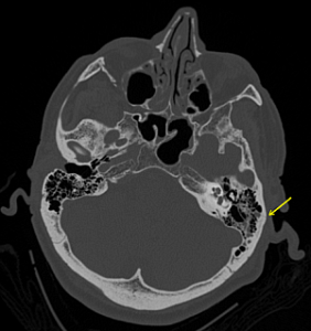
The mastoid portion contains cells or air grooves that can become infected when there is contamination of the middle ear, generating a condition, mainly of pediatric age, called mastoiditis.
Inferior to the squamous portion, it is a curved area that forms the anterior limit of the mastoid process. Its upper face is concave and constitutes the posterior wall of the internal auditory canal..
Its lower face is flatter and is in contact with the mandibular portion of the parotid gland.
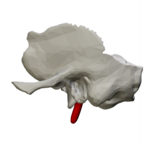
At its lower edge it has a protrusion that projects previously called the styloid process. This bony projection is located just below the ear and serves as an insertion point for various muscles of the tongue and larynx..
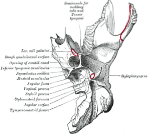
It is a complex part in the shape of a pyramid with the vertex towards the inner side. It contains both some of the most important structures of the middle ear and vital vascular structures, which pass through this portion through specific orifices for each one..
It is widely used for the study of ancient corpses since it usually preserves important traces of DNA that cannot be found in other skeletal remains..
The temporal bone is related to important bone structures that perform protective functions for the structures that contain.
Through the occipito-mastoid suture, it articulates posteriorly with the occipital bone. Below it articulates with the parietal bone. In its squamous portion, it is laterally related to the sphenoid.
The zygomatic process of the temporal bone articulates with the temporal process of the zygomatic bone of the face, forming a structure called the zygomatic arch.
Finally, the temporal bone articulates with the vertical branch of the mandible through the mandibular fossa, forming the temporomandibular joint..
Temporal bone fractures present a high rate of complications for the patient, in addition to being life threatening.
Whenever a polytrauma patient has damage to the skull, the integrity of the temporal bone should be evaluated.
Some of the clinical signs that indicate its fracture are hemorrhagic otorrhea or blood leakage through the ear, vertigo, abnormal movement of the eyes, ringing in the ears or tinnitus and Battle's sign, which is the hematoma over the mastoid process, among others..
However, the absence of these signs does not rule out bone injury, so imaging evaluations with computerized axial tomography (CT) should be made, preferably with three-dimensional reconstruction of the structures..
Regarding tumor processes, both benign and malignant, they are rare conditions but they must be taken into account since they can represent an alteration to the patient's quality of life, especially in their hearing.
In children, middle ear infections can contaminate the mastoid cells generating a condition called mastoiditis, which is relatively common..
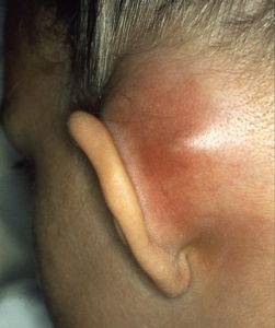
Mastoiditis is difficult to eliminate, requiring prolonged treatment with strong antibiotics. When the infectious process does not respond to conservative treatment, the patient must be operated on to drain the fluid and clean the bone..
This condition is very serious and must be treated promptly as it can progress to affect the protective layers of the brain and the brain itself, forming abscesses..


Yet No Comments