The inotropism is a medical term that refers to the ability of the heart pump to contract. Together with chronotropism, dromotropism and bathmotropism, it forms the 4 fundamental properties of the heart from a functional point of view.
The etymological origin of the word has 3 components of ancient Greek. Ius, which means "nerve" or "fiber"; Tropos, which means "change", "turn" or "turn" and finally -ism, a noun-forming suffix widely used in languages with Latin roots. It would literally translate "change in the fibers" that was adapted to "contraction".

Although the use of the term is reserved almost exclusively to refer to the heart, there is nothing in the medical literature to confirm this. Inotropism could be applied to any muscle in the body and in fact it was so in classical publications, but the current authors stopped doing it. Inotropism outside the heart is not understood today.
Inotropism, like any other property of the heart, can be altered. Although they are not always symptomatic, if the patient who suffers from them presents signs of heart failure, they should receive treatment, which will almost always be aimed at improving or increasing the contractile capacity of the heart..
Article index
When contraction of the heart occurs, all muscle fibers must be activated and the only mechanisms that can modify force generation are changes in fiber length or preload (length-dependent activation) and changes in inotropism (activation independent of length).
The contraction of cardiac muscle fibers basically depends on the intracellular availability of calcium ions. There are other regulatory mechanisms in cardiac inotropism, which will be mentioned later, but it is the calcium concentration that is the most important in a non-pathological setting..
Most of the regulatory pathways for inotropism definitely involve calcium. There are three basic ways through which this cation can positively modify cardiac contraction:
- Increasing its flow during the action potential (mainly during phase 2 of the action potential).
- Increasing its release by the sacroplasmic reticulum (main intracellular calcium store).
- Sensitizing to Troponin-C.
These three effects of calcium favor cardiac contractility, but also limit its duration. By closing the calcium channels of the cell cytoplasm and the sarcoplasmic reticulum, thanks to the activation of potassium channels, the action potential ceases suddenly and intracellular calcium is eliminated in a short time.
This process is repeated cyclically with each heartbeat. This constant inflow and outflow of calcium, with the activation of the sodium and potassium channels, ensures effective cardiac contraction..
The integrity of the myocardial fiber is another of the fundamental elements on which inotropism depends. If there is damage to the muscle fibers of the heart that compromises preload, the amount of calcium available will not matter, the beat will never be completely effective and there will be alterations in pump function.
The preload depends on the length and strain of the cardiac fiber. This phenomenon is governed by the Frank-Starling law which reads: "The contraction energy of the ventricle depends on the initial length of the myocardial fibers ". This means that the more stretched the myocardial fiber is at the end of diastole, the greater the force of contraction..
In short, the myocardial fiber behaves like a spring. The more the spring or myocardial fiber is stretched as the heart fills with blood, the more powerful the force unleashed when the spring is released, that is, the contraction. But if the spring is broken, or the fiber damaged, the energy will be insufficient to generate an efficient heartbeat..
Although they play a minor role, the integrity of the atrioventricular valves is very important to achieve adequate contraction of the heart..
Their closure during the first phase of systole causes the increase in intraventricular pressure necessary to distend the cardiac fiber and produce a correct contraction..
This means that if the valves are damaged or diseased, the ventricle does not fill properly due to pathological return of blood to the atria, the cardiac fiber is not distended, and the energy released does not trigger the contractile force necessary for a normal heartbeat..
Although there is currently no specific method to calculate inotropism, there are indirect ways to do it. The ejection fraction, measured through echocardiography or catheterization, it is a good technique to clinically infer the quality of cardiac contraction.
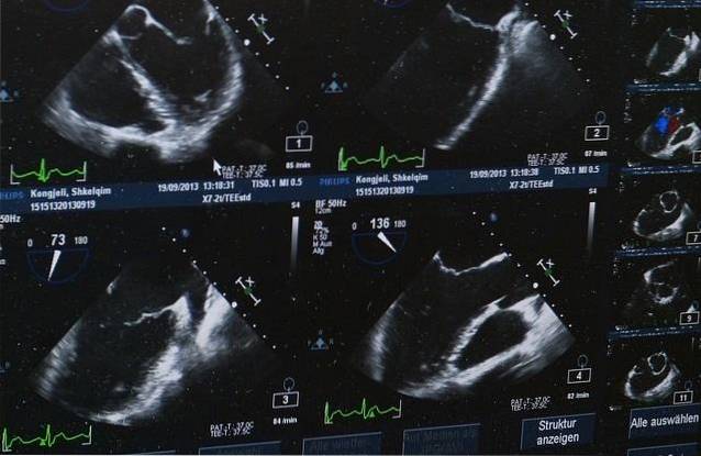
The utility of echocardiography is somewhat broader. It allows estimating (without absolute certainty) the shortening pressure and the increase in pressure / time, both complex but valuable parameters when evaluating the contractility of the heart..
The activity of the atrioventricular valves can also be evaluated through echocardiography..
Any pathologic change in inotropism can lead to heart failure. The same goes for the other three basic functional properties of the heart..
Therefore, in the face of any clinical picture compatible with said disease, a global evaluation must be carried out to determine the level of failure..
Considering the physiology of inotropism, calcium disturbances are some of the most important causes of contractile abnormality. High or low calcium levels can affect heart function. Studies of the myocardium in patients with heart failure have shown failures in the use of cytosolic calcium and in the potency of myocytes.
Diseased myocardial fibers also alter the contractility of the heart. Many people after a myocardial infarction with extensive tissue damage suffer from heart failure due to damage to the muscle fibers.
Chronic hypertensive patients and chagasic patients lose heart muscle compliance and therefore decrease in contractile force.
Some commonly used drugs can compromise cardiac inotropism. Calcium channel blockers, widely used in the treatment of arterial hypertension, have a negative inotropic effect. The same scenario occurs with beta-blockers and most antiarrhythmics..


Yet No Comments