The integrins they are a large group or family of cell surface proteins, apparently unique to the animal kingdom. They are the main resource of cells to maintain interaction (in the form of adhesion) with other cells and with the cell matrix.
Its structure is made up of two subunits called alpha and beta. In mammals it is known that there are between 16-18 alpha units and 3-8 betas, which will act depending on their combination, and also on the physiological state of the cell or specific tissue.
There are several proteins that have adhesive functions. However, the group of integrins is the one that is most distributed and interacts with all the key proteins of the cell matrix. Integrins participate in phagocytosis, cell migration, and wound healing, and are even highly studied for their participation in metastasis.
Article index
They are proteins that are characterized by mechanically joining the cellular cytoskeleton of one cell to another and / or to the extracellular matrix (in a cell-cell and / or cell-matrix interaction). Biochemically, they detect whether or not adhesion has taken place, and transduce cellular signals linking the extracellular environment with the intracellular one, in both directions..
They work or function with other receptors such as immunoglobillins, cadherin, selectins, and syndecands. Regarding the ligands of the integrins, these are constituted by fibronectin, fibrinogen, collagen and vitronectin, among others..
The binding of these to their ligands is due to extracellular divalent cations such as calcium or magnesium. The use of one or the other will depend on the specific integrin.
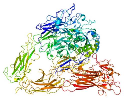
Integrins have an elongated shape ending in a globe-shaped head, which, according to electron microscopy observations, projects more than 20 nanometers from the lipid bilayer.
Integrins are heterodimers, that is, they are molecules always made up of two proteins. Both proteins are considered subunits or protomers and are differentiated as alpha subunit and beta subunit. Both subunits are non-covalently linked. They have a molecular mass of between 90 to 160 kDa.
The number of alpha and beta subunits varies between different groups of organisms in the animal kingdom. In insects such as the fruit fly (Drosophyla), for example, there are 5 alpha and 2 beta subunits, while in nematode worms of the genus Caenorhabditis there are 2 alphas and a beta.
In mammals, the researchers suggest that there is a fixed number of subunits and combinations of these; however, there is no consensus in the literature regarding this number. For example, some mention that there are 18 alpha subunits, 8 beta and 24 combinations, while others speak of 16 alpha and 8 beta for 22 combinations.
Each subunit has the following structure.
The alpha subunit has a structure with a β-helix domain of seven sheets or sheets that form the head, a domain in the thigh, two domains of the calf, a single transmembrane domain and also a short cytoplasmic tail that does not present enzymatic activity or actin binding.
It presents chains with about 1000 to 1200 residues. Can bind divalent cations.
In mammals, which is where the integrins have been studied the most, the alpha subunits can be grouped according to whether or not they contain an inserted domain (alpha I).
The alpha I inserted domain consists of a 200 amino acid region. The presence of this domain in the integrins indicates that they are receptors for collagen and leukocytes..
The alpha integrins that do not have the integrated domain are classified into 4 subfamilies, which we will see below.
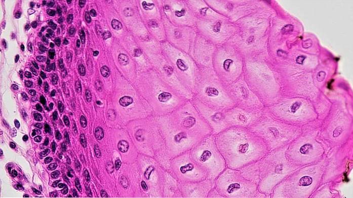
Glycoprotein receptors, also called laminins, are vital for integrating muscle, kidney and skin tissues.
This subfamily is the receptor for arginylglycylaspartic acid, also known as RGD or Arg-Gly-Asp..
This subfamily has been observed in invertebrates, particularly insects. Although little is known about it, there are studies evaluating its essential role in the functional activity of the CD11d leukocyte integrin gene in humans..
This subfamily is known as the alpha 4 / alpha 9 group and comprises the subunits with those same names..
Said subunits are capable of pairing with the beta 1 and beta 7 subunits. They also share ligands very similar to the alpha subunits that present the inserted alpha I domain, such as vascular cell adhesion molecules, blood soluble ligands, fibrinogen and others. including even pathogens.
Structurally, the beta subunit consists of a head, a section called the stem / leg, a transmembrane domain, and a cytoplasmic tail. The head is composed of a beta I domain, which inserts into a hybrid domain that binds to the plexin-semaphore-integrin domain, also known as PSI..
The stem / leg section contains four modules equal or very similar to the cysteine-rich integrin epidermal growth factor and, as already mentioned, a cytoplasmic tail. This cytoplasmic tail, as in the alpha subunit, has no enzymatic or actin-binding activity..
They have chains with a number of residues ranging between 760 and 790, and can bind, like the alpha subunits, bivalent cations.
Integrins have multiple functions, however for which they are mainly known are those that we will see below.
The connection that exists between the cell and the extracellular matrix thanks to the integrins favors the resistance of the cell to mechanical pressure, preventing them from being torn from the matrix.
Several studies suggest that coupling to the cell matrix is a basic requirement for the development of multicellular eukaryotic organisms..
Cell migration is a process in which integrins intervene by binding or coupling to different substrates. Thanks to this they intervene in the immune response and wound healing.
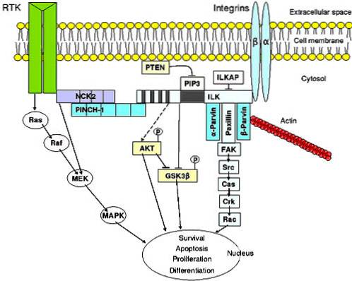
Integrins participate in the signal transduction process. This means that they intervene in the reception of information from the extracellular fluid, they encode it and then the alteration of intracellular molecules begins, as a response.
This signal transduction is involved in a large number of physiological processes such as programmed cell destruction, cell differentiation, meiosis and mitosis (cell division), and cell growth, among others..
Several studies show that integrins play an important role in tumor development, especially in metastasis and angiogenesis. An example of this are the integrins αVβ3 and α1β1, among some others..
These integrins have been related to cancer growth, increased therapeutic resistance, and hematopoietic neoplasms.
An efficient adhesion between cells to form tissues was, without a doubt, a crucial characteristic that must have been present in the evolutionary development of multicellular organisms..
The emergence of the integrin family has been traced back to the appearance of the metazoans around 600 million years ago..
A group of animals with ancestral histological characteristics are the poriferous, commonly called sea sponges. In these animals, cell adhesion occurs by an extracellular proteoglycan matrix. Receptors that bind to this matrix possess a typical integrin-binding motif.
In fact, in this animal group, genes related to specific subunits of some integrins have been identified..
In the course of evolution, the ancestor of the metazoans acquired an integrin and a binding domain to it that has been conserved over time in this immense animal group..
Structurally, we see the maximum complexity of the integrins in the group of vertebrates. There are different integrins that are not present in invertebrates, with new domains. Indeed, more than 24 different functional integrins have been identified in humans - while in fruit flies Drosophila melanogaster there are only 5.


Yet No Comments