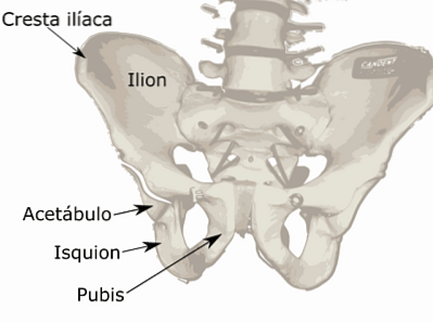
The ischium It is an even bone structure, which is part of the skeleton of the pelvis or bony pelvis. It is found fused with two other bones, the ilium and the pubis. The union of the three pelvic bones is known as hip bone and it is articulated in its posterior part, with the sacrum. This joint is strongly secured by means of firm and resistant ligaments..
In its lower internal part, it articulates with the pubis; in its upper part with the ilium and in its lower external part, it joins with the head of the femur to form the hip joint.
The pelvis is the part of the skeletal system that joins the trunk with the lower limbs. Through its joints with the spine and with the legs, it provides mobility to the body.
The ischium, like the rest of the bones that make up the pelvis, serves as an insertion point for the muscular bodies that make up the pelvic floor. For this reason, it has a fundamental function in the support of the internal organs.
Article index
The ischium is a bone that forms the bony part of the pelvis. It is fused with the other two bones that make it up, above the ilium and below the pubis..
It is an even bone, it is found on both sides of the body. It is a fundamental part of the hip joint, since a large percentage of its body is articulated with the head of the femur.
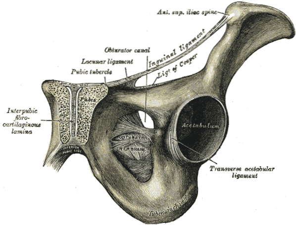
Its structure is very similar to that of the pubis, as it consists of a body, a branch and a tuber. The branch of the pubis and the ischium are united, forming a pelvic foramen called plug hole, through which important vascular and neurological structures pass.
The ischium also serves as a supporting structure for many muscles and ligaments that form the so-called pelvic floor, which is a muscular base whose function is to contain the internal organs within the pelvis such as the bladder, rectum and uterus in women.
The first cartilage outlines that will form the skeleton begin to be observed from the fourth week of gestation.
The ischium and ilium are the first bones of the pelvis to differentiate and find their position in the body of the fetus..
By the ninth week, you can already see the slow and progressive formation of these structures.
The pelvic bones begin to fuse by the 12th week. The entire process of joining these bones happens slowly from birth to adolescence..
Between the ages of 15 and 17, the pelvis is fully fused and the muscles are almost fully developed.
Despite being a medium-sized bone, the ischium has a complicated structure due to its multiple projections, concavities, and muscle relationships. It consists of a body, an upper and a lower branch.
In addition to this, it has two prominences in its postero-inferior part that are of great importance for movement.
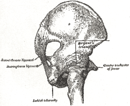
The body is the medial portion of the bone. From its posterior edge a projection called ischial spine. This is where the pelvic muscle originates. upper twin.
It represents an important structure because it forms more than half of the socket where the head of the femur will be installed to form the hip joint. This area is called acetabulum.
The acetabular fossa is formed by the three bones of the pelvis, but the largest surface is provided by the ischium.
The superior or descending branch is a cubic surface in which some of the important muscles of the pelvic floor originate, such as the quadratus femoris muscle, the transverse perineal muscle and the ischiocavernosus..
The lower or ascending branch, for its part, is the thinnest and flattest part of the bone. It is usually called ischiopubic branch, because in its anterior part it meets the lower branch of the pubis and together they form the plug hole.
The obturator foramen serves as a passageway for important vascular and neurological elements that nourish the pelvis and upper thigh..
Its surface is also the origin of various muscles of the pelvic floor, such as the internal obturator, adductor magnus and transverse perineum..
Both branches connect with the upper part of the femur through ligaments that go from this bone to insert into the projections of the other. In this way, the pelvis is connected to the lower limbs through the hip joint..
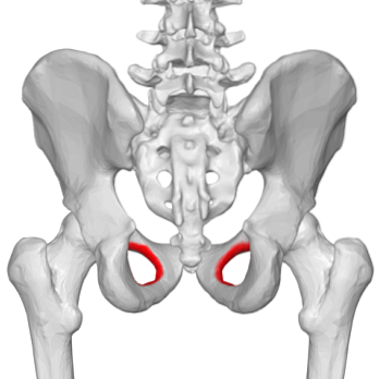
Ischial tuberosity or ischial tuberosity is a robust and irregular convexity found in the posterior and upper part of the lower branch of each ischium. A smoother upper part and a rustic lower part are recognized..
These bony protrusions can be easily palpated with the patient in the fetal position, over the mid-buttock, at the same level as the hip..
They have a mechanical and an anatomical function. From them originate the biceps femoris, semitendinosus and semimembranosus muscles, which are the ones that form the back of the thigh.
The origin of these muscles in this area, make the ischial tuberosities a fundamental element for sitting.
In topographic anatomy, the union, through an imaginary line, of both ischial tuberosities is used as a limit to separate the pelvic floor anteriorly and posteriorly..
This allows the precise description of injuries and is also a guide to recognize, during surgery, the anatomical elements that are related to them.
The ischium is one of the bones that are fused to form the bony pelvis or pelvic girdle.
Given that it is rich in blood vessels, and due to its important relationships with nearby muscles and neurological structures, the surgeon who operates this area must be fully aware of the anatomy of the region.
The obturator artery, which is a branch of the iliac that comes directly from the aorta, makes its way through the obturator foramen. This is accompanied by the nerve and the homonymous vein.
These elements nourish the lower limbs, providing branches that mainly benefit the gluteal, pelvic and upper femur muscles..


Yet No Comments