
The Nervous System is not only made up of neurons. Along with neurons, which are the functional unit of the NS, we find glial cells (neuroglia or glia).
The glia, also called glial cells, are cells of the nervous system. They are part of a support system and are essential for the proper functioning of the nervous system tissue. Unlike neurons, glial cells do not have axons, dendrites, or nerve conduits. Neuroglia are smaller than neurons and are approximately three times more numerous in the nervous system.
They are also much more abundant than neurons; there are ten to fifty times more glial cells than neurons in the vertebrate CNS. Glial cells were described around 1850 by Rudolf Virchow (1821 to 1902).
Contents
The word glía means 'tail' in Greek. Thus, the term neuroglia would mean "adhesive of neurons". This name was given by Rudolf Virchow because he thought that these cells served as an adhesive for neurons, which joined them to form nervous tissue. Thus, the main function of glial cells would be structural, that is, to provide physical support to neurons..
Glia cells are found around neurons and perform very important functions, such as providing structural and metabolic support to neurons..
The set of glial cells is called neuroglia.
There are several types of glial cells present in the central nervous system (CNS) and the peripheral nervous system (PNS) of humans. The six main types of glia include the following:
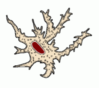
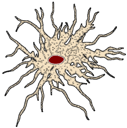
They are the most abundant glial cells and are named in this way because of their stellate shape.
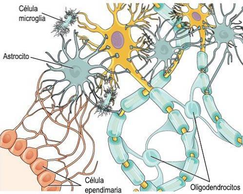
They are found in the brain and spinal cord. They are star-shaped neuroglia that reside in the endothelial cells of the CNS that form the blood-brain barrier. This barrier restricts what substances can enter the brain. Protoplasmic astrocytes are found in the gray matter of the cerebral cortex, while fibrous astrocytes are found in the white matter of the brain. Other functions of astrocytes include glycogen storage, nutrient provision, regulation of ion concentration, and neuron repair..
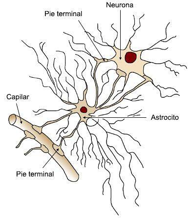
Astrocytes
Ependymal cells are specialized cells that line the brain ventricles and the central canal of the spinal cord. They are found within the choroid plexus of the meninges. These hair cells surround the capillaries of the choroid plexus and form cerebrospinal fluid..
They form the epithelial lining of the ventricles of the brain and the central canal of the spinal cord.
Ependymal cells, like other neuroglial cells, derive from a layer of embryonic tissue known as the neuroectoderm..
They give rise to the epithelial layer that surrounds the choroid plexus in the lateral ventricles of the cerebral hemisphere. These epithelial cells mainly produce cerebrospinal fluid..
Ependymal cells have cilia and are located in front of the cavity of the ventricles. The coordinated movement of these cilia influences the direction of cerebrospinal flow, the distribution of neurotransmitters and other messengers to neurons..
Ependymal cells called Tanicytes play an important role in the transport of hormones in the brain..
Microglia are extremely small cells of the central nervous system that eliminate cellular debris and protect against microorganisms (bacteria, viruses, parasites, etc.). Microglia are thought to be macrophages, a type of white blood cell that protects against foreign matter. They also help reduce inflammation by releasing anti-inflammatory cytokines..
Microglia
Under normal conditions, the number of microglia cells is small, but when injury or inflammation of the nervous tissue occurs, these cells proliferate rapidly (as do astrocytes) and migrate to the area of injury to engulf the remains damaged cells, myelin fragments, or neurons.
Microglia act as a phagocytic cell and protect the brain from invading microorganisms.
Oligodendrocytes are structures in the central nervous system that surround some neuronal axons to form an insulating layer known as the myelin sheath. The myelin sheath, made up of lipids and proteins, functions as an electrical insulator for axons and promotes more efficient conduction of nerve impulses.
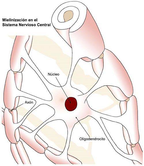
Oligodendrocyte. An oligodendrocyte can myelinate segments of different axons
They form the myelin layer of the CNS: a single oligodendrocyte can myelinize different segments of the same axon or different axons (from 20 to 60 different axons).
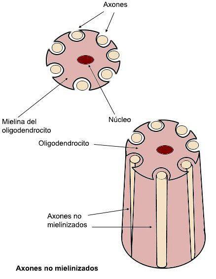
An oligodendrocyte surrounds different unmyelinated axons
The oligodendroglia also has a protective function on the unmyelinated axons, since it surrounds them and keeps them fixed.
Oligodendroglia forms the myelin sheath in the CNS.
There are autoimmune diseases that destroy the myelin layer: in multiple sclerosis the cells that make up myelin are not recognized by the body as their own and are destroyed. This disease is progressive, and depending on the number and function of neurons that lose myelin, the consequences will be more or less serious.
These glial satellite cells cover and protect neurons in the peripheral nervous system. Provide structural and metabolic support for sensory, sympathetic, and parasympathetic nerves.
Astroglia
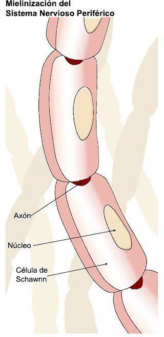
In the PNS, each Schawnn cell forms a single myelin segment for a single axon.
In the peripheral nervous system (PNS), Schawnn cells perform the same functions as the different glial cells of the CNS. These functions are as follows:
Bradford, H.F. (1988). Fundamentals of neurochemistry. Barcelona: Labor.
Carlson, N.R. (1999). Physiology of behavior. Barcelona: Ariel Psychology.
Carpenter, M.B. (1994). Neuroanatomy. Fundamentals. Buenos Aires: Editorial Panamericana.
Delgado, J.M .; Ferrús, A .; Mora, F .; Rubia, F.J. (eds) (1998). Neuroscience Manual. Madrid: Synthesis.
Diamond, M.C .; Scheibel, A.B. and Elson, L.M. (nineteen ninety six). The human brain. Work book. Barcelona: Ariel.
Guyton, A.C. (1994) Anatomy and physiology of the nervous system. Basic neuroscience. Madrid: Editorial Médica Panamericana.
Kandel, E.R .; Shwartz, J.H. and Jessell, T.M. (eds) (1997) Neuroscience and Behavior. Madrid: Prentice Hall.
Martin, J.H. (1998) Neuroanatomy. Madrid: Prentice Hall.
.


Yet No Comments