
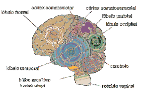
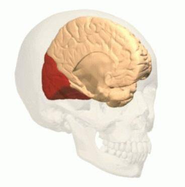
The occipital lobe it is the part of the brain where images are processed. It is one of the smallest cerebral lobes of the cerebral cortex, located in the back of the skull, between the cerebellum, the parietal lobe and the temporal lobe..
When referring to the occipital lobe, it is more convenient to speak of occipital lobes in the plural, since there are two occipital structures, one in each hemisphere of the brain.
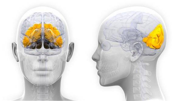
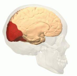
The two occipital lobes that human beings have are practically symmetrical and the main function of both lies in the processing of visual information. The occipital region is characterized by being one of the smallest lobes of the cortex and is located at the back of the brain, just above the nape..
Article index
The occipital lobe is divided into two cerebral hemispheres. Therefore, each brain contains a right occipital lobe and a left occipital lobe, which are separated by a narrow fissure..
Evolutionarily, the occipital lobe stands out for not having undergone excess growth throughout the evolution of the species. Unlike other brain regions that have increased in size throughout the evolution of the ancestors, the occipital lobe has always presented a similar structure.
This means that while other regions of the human cerebral cortex have developed and organized in a more complex way, the occipital lobe has remained with similar structures for the last hundreds of thousands of years..
On the other hand, the occipital lobe is characterized by not being especially vulnerable to injury, since it is located in the posterior region of the brain. However, severe trauma to this brain region usually generates modifications in the visual-perceptual system.
The occipital lobe acts as an area of reception and visual integration, picking up signals that come from different regions of the brain. Anatomically, it constitutes one-eighth of the cerebral cortex and contains primary visual and visual association areas.
In general, the occipital lobe can be divided into two large structures: the primary visual cortex and the areas of visual association..
Despite the fact that this anatomical division of the occipital lobe allows a better description of its structure and functioning, in practice the anatomical limits between both structures tend to be less identifiable..
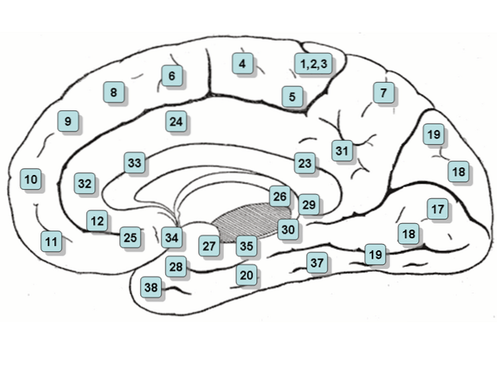
The area of the primary or striated visual cortex (Brodman's area 17) is located in the convolutions that originate the walls of the calcarian fissure and is characterized by receiving optical radiation.
The lower half of the contralateral field of vision is represented on the upper wall of the calcarial fissure (wedge). The upper half of the contralateral visual field is represented on the lower wall of the calcarial fissure (lingual gyrus)..
Finally, in the posterior half of the primary visual cortex is macular vision. Generally, unilateral lesions in this area of the occipital lobe produce a contralateral homonymous hemianopia..
The visual association areas of the occipital lobe are formed by the paraestriate areas and the periestriate areas, or what is the same, the areas 18 and 19 of Brodman.
The periestriate area is larger than the paraestriate and forms the largest lateral surface of the occipital lobe.
Brodman's areas 18 and 19 receive visual information from bilaterally striated areas. They are essential regions when it comes to constituting complex visual perceptions related to color, the direction of objects or movement.
Lesions originating in these areas usually cause visual agnosia, that is, an inability to recognize objects and colors.
In order to describe and understand the function of the occipital lobe, it must be taken into account that the different regions that make up the cerebral cortex do not have a single activity. In fact, the different lobes of the cortex participate differently in multiple brain activities.
Despite this factor that defines the functioning of the upper regions of the brain, the function that best describes the activity of the occipital lobe is the processing of visual information.
In fact, the main function of this region of the cortex is to receive the stimuli related to the optic pathway, which come in the first instance from the optic nerves and, in the second instance, from other subcortical structures..
In this sense, the occipital lobe comprises the visual cortex, which is the area of the brain's cortex that is first received by information from the retinas of the eyes and the optic nerves..
Likewise, the visual cortex of the occipital lobe is divided into different regions that are classified according to the level of processing they take care of..
Thus, the primary visual cortex is the part of the occipital lobe that is responsible for processing the "raw" visual data and is the region responsible for detecting the general patterns that can be found in the visual information collected by the eyes..
The general data collected by the primary visual cortex of the occipital lobe are usually not very detailed and do not usually contain specific information about the stimulus captured.
Subsequently, the primary visual cortex is responsible for sending the collected information to other regions of the occipital lobe, which are responsible for carrying out a more refined processing of vision..
Likewise, the other structures of the occipital lobe are responsible for sending the analyzed information to other structures of the brain..
In summary, the occipital lobe contains the areas or nerve centers that mainly regulate the following activities:
The occipital lobe has two main routes of communication with other regions of the brain. These pathways allow the transmission of the information that reaches the primary visual cortex and, therefore, send the visual information to the corresponding brain structures..
The dorsal pathway of the occipital lobe is responsible for connecting the primary visual cortex with the frontal region of the cerebral cortex. This connection is made through neural networks that are close to the upper region of the skull..
In this way, through this pathway, the information processed by the primary visual cortex reaches the parietal lobe through the third and fifth visual cortex..
This processing pathway of the occipital lobe is responsible for establishing the characteristics of the location and movement of visual stimuli. For this reason, the dorsal pathway is also known as the "where" pathway and the "how" pathway, as it allows us to elaborate and examine these elements of visual stimuli..
The ventral pathway of the occipital lobe starts from the primary visual cortex and goes to the frontal region of the brain through the lower part of the brain. That is, it adopts a similar route to that of the dorsal pathway but passes through the lower regions of the cortex..
This pathway is carried out through the second and fourth visual cortex and is responsible for processing the information collected and analyzed by the primary visual cortex..
The neural network that constitutes this transmission path is in charge of processing the characteristics of the isolated elements that are being visualized at all times..
That is, the ventral pathway of the occipital lobe allows information about the content of visual stimuli to be transmitted to other brain areas. For this reason, this road is also known as the "what" road..
The occipital lobe is one of the regions of the brain that experiences the least damage. Being located in the back of the brain, it is quite protected from pathologies.
However, the trauma suffered in this area of the skull can produce subtle modifications in the functioning of the occipital lobe, a fact that can translate into visual-perceptual distortions. In fact, the damage suffered in this lobe usually causes defects and scatomas in the field of vision..
More specifically, lesions originating in the Peristriate region of the occipital lobe (a structure involved in visual spatial processing) often generate alterations in movement and color discrimination.
On the other hand, certain damage to the occipital lobe can cause a homonymous loss of vision with exactly the same field cut within both eyes..
Research has shown that occipital lobe disorders can lead to hallucinations and perceptual illusions. These can be caused both by injuries in the occipital region and by temporal seizures of the lobe.
Visual illusions (disturbances in perception) can take the form of objects that appear larger or smaller than they really are, objects that lack color, or objects that are abnormally colored.
Finally, lesions in the parietal-temporal-occipital area of the association can cause speech blindness with impairments of writing.
Recent studies have shown that the occipital lobe could be a very important brain structure in the development of epilepsy.
Although nowadays there are still no irrefutable data, many authors point out that the occipital lobe would have a prominent role in the appearance of epileptic seizures, or at least in part of them.
In this sense, occipital lobe epilepsies have been described, which are characterized by being simple partial seizures or secondarily generalized.
The clinical manifestations of this condition usually include, but not always, visual symptoms and are often associated with migraine..
In occipital lobe epilepsy, simple negative visual manifestations such as scatoms (spots in the field of vision), hemianopsia (blindness of one area of the field of vision) or amaurosis (blindness) can occur..
Likewise, in some cases it can also generate simple positive manifestations such as phosphenes (flashes of light), flashes or sparks.
The visual sensations of occipital lobe epilepsy are usually manifested in the visual field contralateral to the occipital cortex in which the discharge develops. However, in some cases the sensations can spread and compromise all visual fields.
In occipital lobe epilepsy, alterations in perception have also been described, such as: increase in the size of objects or images, decrease in objects or images, and changes in shape..
In some rare cases, perceptual alterations can be highly complex and the person can view entire scenes as if “a movie was playing in the head”.
In other rare cases, occipital lobe epilepsy can cause autoscopy (the person perceives how he is observing himself as if he were an outside observer).
These manifestations are very hallucinatory and are usually located preferably in the area where the temporal, parietal and occipital lobes converge.
Finally, the motor seizures of this type of condition usually include a deviation of the head and eyes to the opposite side of the hemisphere where the epileptic discharge occurs..
The discharge can extend towards the temporal or parietal lobes, and in some cases it can even reach the frontal lobe. Sometimes it spreads to the occipital cortex of the opposite hemisphere and can become generalized encompassing the entire cortex.


Yet No Comments