The lactate dehydrogenase, Lactic acid dehydrogenase, NAD-dependent lactate dehydrogenase or simply LDH, is an enzyme belonging to the group of oxidoreductases that is found in practically all animal and plant tissues and in many microorganisms such as bacteria, yeasts and archaea.
Enzymes of this type are denoted by number EC 1.1.1.27 of the Enzyme Nomenclature Committee and are responsible for the reaction that converts lactate to pyruvate (by oxidation) and vice versa (by reduction), oxidizing or reducing nicotinamide adenine dinucleotides ( NAD + and NADH) in the process known as lactic fermentation.
Unlike alcoholic fermentation, which occurs only in some microorganisms such as yeast and uses glycolytic pyruvate for the production of ethanol, lactic fermentation takes place in many organisms and body tissues of different living beings.
This important enzyme for cell metabolism was crystallized from rat skeletal muscle in the 1940s and, to date, the best characterized are skeletal muscle and mammalian cardiac tissue..
In "higher" animals the enzyme uses the L isomer of lactate (L-lactate) for the production of pyruvate, but some "lower" animals and bacteria produce D-lactate from pyruvate obtained by glycolysis..
Lactate dehydrogenase is usually expressed mainly in tissues or cells under anaerobic conditions (with low blood supply) that, in humans, for example, can characterize pathological conditions such as cancer, liver or heart conditions.
However, the conversion of pyruvate to lactate is typical of the muscles during exercise and the cornea of the eye, which is poorly oxygenated.
Article index
Lactate dehydrogenase serves multiple functions in numerous metabolic pathways. It is the center of the delicate balance between the catabolic and anabolic carbohydrate pathways.
During aerobic glycolysis, pyruvate (the last product in the pathway per se) can be used as a substrate for the pyruvate dehydrogenase enzyme complex, by which it is decarboxylated, releasing acetyl-CoA molecules that are used downstream, metabolically speaking, in the Krebs cycle.
In anaerobic glycolysis, on the other hand, the last step of glycolysis produces pyruvate, but this is used by lactate dehydrogenase to produce lactate and NAD.+, that restores the NAD+ that was used during the reaction catalyzed by glyceraldehyde 3-phosphate dehydrogenase.
As during anaerobiosis the main source of energy production in the form of ATP is glycolysis, lactate dehydrogenase plays a fundamental role in the reoxidation of NADH produced in previous steps of the glycolytic pathway, essential for the functioning of other related enzymes.
Lactate dehydrogenase is also involved in glycogenesis that takes place in tissues that convert lactate to glycogen and, in some aerobic tissues such as the heart, lactate is a fuel that is reoxidized to produce energy and reducing power in the form of ATP and NAD+, respectively.
There are multiple molecular forms of lactate dehydrogenase in nature. Only in animals it has been determined that there are five lactate dehydrogenase activities, all tetrameric and essentially composed of two types of polypeptide chains known as the H and M subunits (which can be homo- or heterotetrameric).
The H form is typically found in cardiac tissue, while the M form has been detected in skeletal muscle. Both chains differ from each other in terms of abundance, amino acid composition, kinetic properties, and structural properties..
The H and M forms are the translational product of different genes, possibly located on different chromosomes, and which are also under the control or regulation of different genes. The H form is predominant in tissues with aerobic metabolism and the M form in anaerobic tissues..
Another type of nomenclature uses the letters A, B and C for the different types of enzymes in both mammals and birds. Thus, muscle lactate dehydrogenase is known as A4, the cardiac as B4 and a third is called C4, which is specific to the testicles.
The expression of these isoenzymes is regulated both in a developmental-dependent and tissue-dependent stages..
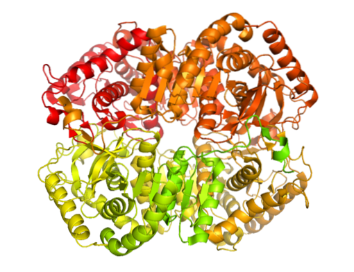
The enzyme has been isolated from different animal sources and it has been determined that its tetrameric structure has an average molecular weight of about 140 kDa and that the binding site for NADH or in NAD+ consists of a β-folded sheet composed of six chains and 4 alpha helices.
Lactate dehydrogenase activity of animal origin is determined spectrophotometrically in vitro by color change measurements due to the redox process that takes place during the pyruvate to lactate conversion reaction.
Measurements are made at 340nm with a spectrophotometer and the rate of decrease in optical density due to the oxidation or "disappearance" of NADH, which is converted to NAD, is determined.+.
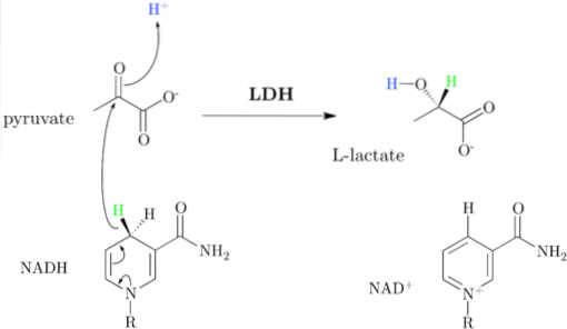
That is, the determined reaction is as follows:
Pyruvate + NADH + H+ → Lactate + NAD+
The enzymatic measurement must be carried out in optimal conditions of pH and concentration of substrates for the enzyme, so that there is no risk of underestimating the amount present in the samples due to a deficit of substrates or due to extreme conditions of acidity or basicity..
Another method, perhaps a bit more modern, for determining the presence of lactate dehydrogenase has to do with the use of immunological tools, that is, with the use of antibodies..
These methods take advantage of the affinity between the binding of an antigen with an antibody specifically generated against it and are very useful for the rapid determination of the presence or absence of enzymes such as LDH in a particular tissue..
Depending on the purpose, the antibodies used must be specific for the detection of any of the isoenzymes or for any protein with lactate dehydrogenase activity..
The determination of this enzyme is carried out for different purposes, but mainly for the clinical diagnosis of some conditions, among which myocardial infarction and cancer stand out..
At the cellular level, the release of lactate dehydrogenase has been considered as one of the parameters to determine the occurrence of necrotic or apoptotic processes, since the plasma membrane becomes permeable.
The products of the reaction that it catalyzes can also be determined in a tissue in order to determine whether an anaerobic metabolism predominates for any particular reason..
As initially mentioned, the enzyme lactate dehydrogenase, whose systematic name is (S) -lactate: NAD+ dehydrogenase, catalyzes the conversion of lactate to pyruvate in the NAD form+ dependent, or vice versa, which occurs thanks to the transfer of a hydride ion (H-) from pyruvate to lactate or from NADH to oxidized pyruvate.
The NAD+ It has a unit of ADP and another nucleotide group derived from nicotinic acid, also called niacin or vitamin B3, and this coenzyme participates in multiple reactions of great biological importance.
It is important to note that the equilibrium in this reaction is shifted towards the side corresponding to lactate and it has been shown that the enzyme is also capable of oxidizing other acids (S) -2-hydroxymonocarboxylics and employ, although less efficiently, NADP+ as a substrate.
Depending on the body region under consideration and, at the same time, on its metabolic characteristics in relation to the presence or absence of oxygen, tissues produce different amounts of lactate, the product of the reaction catalyzed by LDH..
Considering, for example, a red blood cell (erythrocyte) that lacks mitochondria that can metabolize pyruvate produced during glycolysis to COtwo and water, then it could be said that these are the main lactate-producing cells in the human body, since all pyruvate is converted to lactate by the action of lactate dehydrogenase.
On the other hand, if liver cells and skeletal muscle cells are considered, they are responsible for the production of a minimum amount of lactate, since it is rapidly metabolized..
The concentration of lactate dehydrogenase in the blood serum is the product of the expression of several isoenzymes in the liver, heart, skeletal muscle, erythrocytes, and tumors, among others..
In blood serum, the normal ranges of lactate dehydrogenase activity are between 260 and 850 U / ml (units per milliliter), with an average value of 470 ± 130 U / ml. Meanwhile, blood hemolysates have an LDH activity that varies between 16,000 and 67,000 U / ml, which is equivalent to an average of 34,000 ± 12,000 U / ml..
The quantification of the lactate dehydrogenase concentration in the blood serum has an important value in the diagnosis of some heart diseases, liver, blood and even cancers.
High levels of LDH activity have been found in patients with myocardial infarctions (both experimental and clinical), as well as in cancer patients, specifically in women with endometrial, ovarian, breast and uterine cancer.
Depending on the particular isozyme found in "excess" or high concentration, quantification of lactate dehydrogenase isoenzymes is used by many treating physicians for the determination of tissue damage (severe or chronic)..


Yet No Comments