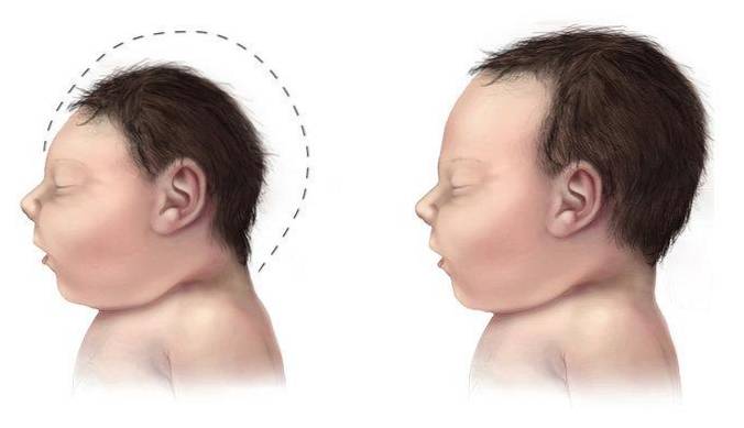
The macrocephaly it is a neurological disorder in which there is an abnormal increase in the size of the head. Specifically, there is an increase in the cranial perimeter, that is, the distance around the widest or upper area of the skull is greater than expected for the age and sex of the affected person.
At a more clinical level, macrocephaly occurs when the head circumference or perimeter is above the mean for that age and gender by 2 standard deviations or is greater than the 98th percentile. These signs may be evident from birth or develop in the early life.
In general, it is a rare disorder that affects men more often than women. Although not all cases of macrocephaly are cause for alarm, it is often accompanied by various symptoms or medical signs: generalized developmental delay, seizures, corticospinal dysfunctions, among others..
Article index
Macrocephaly is a neurological disorder that is included within the cranial growth disorders.
In cranial growth pathologies or disorders, cranial size abnormalities occur due to different alterations in the bones of the cranial vault or in the central nervous system.
Specifically, macrocephaly is defined as an abnormal increase in the cranial perimeter that is above the expected value for the age and sex of the affected person (García Peñas and Romero Andújar, 2007).
This type of alterations can be due to an excess volume of cerebrospinal fluid, an increase in the size of the brain or even a
thickening of the cranial vault.
Although a large number of those affected by macrocephaly do not present significant signs or symptoms derived from the pathology, many others present significant neurological abnormalities.
There are no specific statistical data on the prevalence of macrocephaly in the general population..
However, clinical studies consider it to be a rare or infrequent pathology, which occurs in approximately 5% of the population.
(Mallea Escobar et al., 2014).
It is generally a disorder that affects the male sex in a greater proportion and is usually present already at birth or develops in the first
years of life, therefore infantile macrocephaly is common.
Derived from the definition of this pathology, the most characteristic symptom of macrocephaly is the presence of an abnormally large head size.
As in the case of other pathologies or disorders that affect cranial growth, the size of the head is measured through the circumference or cranial perimeter, the measurement of the contour of the head from the top (Microcephaly, 2016).
The size of the head or skull is determined both by the growth of the brain, the volume of cerebrospinal fluid (CSF) or blood, and by the bone thickness of the skull (Mallea Escobar et al., 2014).
A variation in any of these factors may cause important neurological consequences, therefore it is crucial that a control and measurement of the growth of the cranial perimeter is carried out in newborns and children, especially during the first years of life (Mallea Escobar et al., 2014).
The standard growth patterns show us the following values (Mallea Escobar et al., 2014):
- Head circumference in term newborns: 35-36cm.
- Approximate growth of the head circumference during the first year of life: approximately 12cm, more accentuated in males.
- Speed of increase in the head circumference during the first three months of life: about 2cm per month.
- Speed of increase in the head circumference during the second trimester of life: about 1cm per month.
- Rate of increase in the head circumference during the third and fourth trimesters of life: about 0.5cm per month.
The values obtained from the measurement of the head size in the medical and health controls should be compared with a standard or expected growth chart. Children with macrocephaly present values significantly higher than the average for their age and sex.
Due to the different etiologies that will give rise to the increase in the size of the head, different medical complications may appear that affect both the neurological functioning and the general functional level of the affected person..
The medical conditions associated with macrocephaly will depend on the etiological cause, despite this, there are some frequent clinical manifestations (Martí Herrero and Cabrera López, 2008):
- Asymptomatic macrocephaly.
- Convulsive episodes.
- Generalized delay in development, cognitive and intellectual deficits, hemiparesis, etc..
- Vomiting, nausea, headaches, drowsiness, irritability, poor appetite.
- Gait disturbances and deficits, visual deficits.
- Signs of intracranial hypertension, anemia, biochemical alterations, systemic bone pathologies.
As we pointed out previously, macrocephaly may appear due to different alterations that affect the size of the brain, volume of cerebrospinal fluid or due to bone abnormalities..
One of the publications of the Spanish Association of Pediatrics About macrocephaly and microcephaly, he makes a detailed classification of the possible etiological causes of macrocephaly (Martí Herrero and Cabrera López, 2008):
In the case of macrocephaly due to the presence or development of a brain and / or cerebrospinal fluid pathology, macrocephaly of primary or secondary origin may be found..
Primary microcephaly occurs as a consequence of an increase in the size and weight of the brain.
Generally, in this type of microcephaly a greater number of nerve cells or a greater size can be observed. When the presence of this etiological cause is determined, the pathology is called macroencephaly.
This type of alterations usually have a genetic origin and therefore, familial macrocephaly and hemimegalencephaly form part of this classification.
In addition, it is common for macroencephaly to form the set of clinical manifestations of other pathologies such as: bone dysplasias, fragile X, Sotos syndrome, Beckwith syndrome, chromosomopathies, etc..
Secondary microcephaly, also called progressive or evolutive microcephaly, may be due to alterations in the volume of cerebrospinal fluid, the presence of lesions or the presence of occupying substances.
- Increased level and volume of cerebrospinal fluid (CSF): abnormalities in the production, drainage or reabsorption of cerebrospinal fluid can cause an accumulation of this and therefore lead to Hydrocephalus.
- Presence of occupant injuries: this type of alterations refer to the presence of intracerebral structural and vascular malformations, masses or collections. Some of the pathologies that give rise to this type of injury are: cysts, tumors, bruises, arteriovenous malformations, etc..
- Presence of abnormal substances: these types of alterations refer to the presence of deposit or metabolic diseases such as Alexander's disease, Canavan's disease, metabolic diseases, etc..
As for the cases of macrocephaly that are due to bone abnormalities, we can find:
- Macrocephaly due to early closure of cranial sutures.
- Macrocephaly due to systemic bone abnormalities: rickets, osteogenesis, osteoporosis, etc..
Macrocephaly is a neurological pathology that can be detected during the gestation phase.
Routine health checks through ultrasound ultrasound are capable of detecting abnormalities in cranial growth during the early stages of pregnancy, when macrocephaly has a congenital or prenatal origin..
However, it is not always possible to detect it before birth, since many cases of macrocephaly occur secondary to other medical conditions.
It is usually detected in pediatric consultations through the measurement of the cranial perimeter. In addition, different neurological analyzes must also be performed to determine the etiological cause..
Specifically, the clinical examination must include (Martí Herrero and Cabrera López, 2008):
- Physical examination of the skull- An accurate measurement of the head circumference and a comparison with growth standards should be made.
- Neurological examination: it will also be necessary to evaluate different neurological factors (gait, motor coordination, sensory deficits, cerebellar signs, reflexes, etc.).
- Pediatric examination: in this case, it will be oriented towards the study of the etiological cause of macrocephaly through the analysis of genetic and neurological pathologies, etc..
- Complementary exams: In addition to the physical and neurological examination, some complementary examinations such as magnetic resonance imaging, computed tomography, X-rays, lumbar puncture, electroencephalography, etc. may be required. Especially in those macrocephaly of undetermined origin.
There is currently no curative treatment for macrocephaly. Generally, the treatment is symptomatic and will depend on the precise diagnosis of the etiology.
After the detection of macrocephaly, it is necessary to determine the underlying cause to design the best therapeutic approach, since in cases in which there is hydrocephalus as the main cause of macrocephaly, it will be necessary to use surgical interventions.
Therefore, the treatment will have a marked palliative value. Pharmacological approaches may be used to control medical complications, as well as non-pharmacological ones for the treatment of neurological and cognitive sequelae..
In all cases of macrocephaly and other types of cranial developmental disorders, it is essential that a neurological and / or neuropsychological examination be performed to examine the level of general functioning: developmental deficits, cognitive functions, language deficits, motor skills, etc. (National Institute of Neurological Disorder and Stroke, 2016).
Some of the non-pharmacological interventions that can be used in symptomatic cases of macrocephaly are (Martí Herrero and Cabrera López, 2008):
- Neuropsychological rehabilitation.
- Early stimulation.
- Special education.
- Occupational therapy.
The prognosis and evolution of this pathology depends fundamentally on the origin and associated symptoms..
In children with benign microcephaly, the absence of symptoms or significant medical complications will allow them to develop all areas normally (Erickson Gabbey, 2014).
However, in many other cases, future prospects will depend on the presence of medical complications (Erickson Gabbey, 2014). In general, children suffering from macrocephaly will present significant generalized developmental delays and therefore will require therapeutic intervention to promote the acquisition of new skills and the achievement of an efficient functional level.


Yet No Comments