Malassezia furfur is a species of yeast fungus, causative agent of superficial mycosis pityriasis versicolor, also called ringworm or tinea versicolor. Its distribution is worldwide, but it is more frequent in tropical and temperate climates..
It represents 5% of mycoses in general and 20% of superficial mycoses. In summer, when it is hotter, endemics increase from 4% to 50%. It has been seen to affect both sexes with a slight predilection in women between the ages of 2 and 90, with an average of 20 to 30 years.
Children are affected by approximately 5 to 12%, in ages 8 to 11. The increase of this fungus from adolescence may be linked to hormonal factors where there is greater production of sebum in the skin.
However, other findings that include the presence of the fungus in babies in countries like Thailand, suggests possible climatic and perhaps genetic factors in the colonization of the skin..
Infection by this fungus has no predilection for races or social strata and is not very important in HIV patients, although it is frequent in patients with other immune deficiencies.
Article index
Malassezia furfur it is found as a commensal in the skin microbiota. It is mainly found in areas with a large number of sebaceous glands, such as the scalp, face, outer ear, chest and back; its presence increases with age, usually at puberty.
If the fungus proliferates more than normal, it goes from being a saprophytic to a pathogen. On the other hand, among the characteristics of Malassezia furfur it is an imperfect fungus, that is, it only has asexual reproduction, therefore they reproduce by blastoconidia.
Likewise, it is a lipophilic fungus, that is, it has a predilection for lipids, which it uses as a carbon source. Cases of systemic infections and septicemia due to contamination of deep-guide vascular catheters have been reported in patients receiving parenteral treatment..
Many of the emulsions used in parenteral treatment are rich in long chain fatty acids. This establishes an ideal environment for the fungus to proliferate and enter the bloodstream..
On the other hand, the species of Malassezia have been recognized as skin colonizers in various animals, including bears, monkeys, pigs, elephants, rhinos, and birds.
Kingdom: Fungi
Phylum: Basidiomycota
Class: Exobasidiomycetes
Order: Malasseziales
Family: Malasseziaceae
Gender: Malassezia
Species: furfur
It is characterized by affecting the superficial layers of the skin, specifically the stratum corneum of the epidermis.
Invasion of the outer layers of the stratum corneum occurs after the conversion of the yeast commensal into a filamentous parasite as a consequence of local immunological alterations..
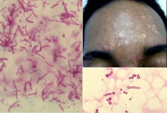
Inflammation and scaling are believed to be the cause or consequence of fungal overpopulation. The fungus causes the appearance of erythematous macules, confluent with hypopigmented and hyperpigmented areas, associated with induration and desquamation.
The lesions are located mainly on the trunk and arms, but can also affect armpits, groin, arms, thighs, buttocks, shoulders, back, neck and face.
They present variable colorations that go from pink to yellow-brownish and are sometimes achromatic. This is where the name versicolor comes from.
Changes in skin color occur by various mechanisms.
On the one hand, the fungus produces dicarboxylic acid, especially azelaic acid and other tyrosinase-dependent lipid metabolites, such as pityriazitrine and pityrialactone, which act on melanocytes and inhibit dopa-tyrosinase. This mechanism manifests with hypochromia.
Whereas, hyperchromic lesions are due to the increase in size of the melanosomes, for which there are two hypotheses:
The infection is usually asymptomatic, but occasionally there may be slight itching and redness of the skin..
If an ultraviolet light is passed to the lesions, they will be observed with a greenish-yellow fluorescent coloration.
The sample is taken with a scalpel, making a scraping, then mounted directly on a sheet with a drop of 20% KOH, plus Parker ink or methylene blue to highlight the structures.
Viewed under the light microscope, the fungus is usually observed as a group of budding yeast cells (in clusters) mixed with short curved hyphae, giving the appearance of spaghetti with meatballs..
Yeasts are oval or bottle-shaped, measuring 3 to 8 µm in diameter. They present with a monopolar bud with a septum in the cell wall where the bud leaves a scar.
Adhesive tape is a very effective method for taking samples of lesions for direct examination. It consists of placing a piece of transparent adhesive tape over the injury, applying pressure to it, and then removing it in the opposite direction to the injury..
The tape is placed on a slide and viewed under a microscope with a 10x to 40x objective. Fixed preparations can also be made from the skin scales.
For sampling with any of the methods it is necessary that the patient has not been treated with fungicides or ointments. In areas subjected to frequent washing such as the face, the direct examination is not very effective.
Differential diagnosis should be made with seborrheic dermatitis, pinta, vitiligo, erythrasma, pityriasis rosea, secondary syphilis, parasitic acromia and circinate ringworm.
The cultivation of the fungus is difficult, therefore it is not usually carried out, since with the previously explained methods the diagnosis can be made.
However, the fungus can grow on Sabouraud's dextrose agar or 5% sheep's blood agar, supplemented with long-chain fatty acids on their surface. For this you can use olive oil.
Malassezia furfur produces smooth, convex creamy colonies with rough variants. At Gram, elongated, spherical or oval cells are observed and some filaments can be visualized.
By electron microscopy, it is possible to see a multilaminar wall, thickened and with diagonal striations. Colonies develop slowly after 2 to 4 days of incubation at 35ºC.
The treatment consists of placing 1% selenium sulfide applied on the lesions every 3 days for 15 minutes, and then washing the area.


Yet No Comments