Rotator cuff characteristics, function, pathologies
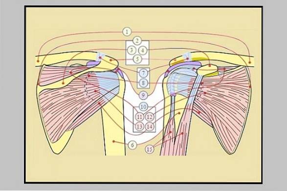
The rotator cuff It is a structural complex formed by four muscles (supraspinatus, infraspinatus, teres minor and subscapularis) and their tendons. These converge on the capsule of the glenohumeral joint, in order to give stability to the joint and coordinate its movements..
The glenohumeral joint has a movement capacity that is not comparable to any other, being capable of executing flexion, extension, adduction, and abduction movements, and as if this were not enough, it also allows internal and external rotation movements..

This great functionality is possible thanks to the anatomical characteristics of the glenoid cavity with respect to the head of the humerus, since it is extremely large for the shallow depth of the glenoid cavity. This of course gives it greater movement capacity, but at the same time makes it more unstable..
The presence of the muscles that make up the rotator cuff is essential to reinforce the union of these two bone structures, although they do so in a secondary way, since there are structures such as the joint capsule, the glenohumeral ligaments and the glenoid rim that act as primary form.
All of these structures, including the rotator cuff, protect and provide stability to the joint, preventing the head of the humerus from slipping out of place. In addition, the rotator cuff together with the deltoid makes upper limb movements possible..
It should be noted that the rotator cuff very frequently suffers alterations that affect the functionality of the shoulder, causing pain.
Article index
- 1 Features
- 1.1 The supraspinatus muscle
- 1.2 Infraspinatus muscle
- 1.3 Teres minor muscle
- 1.4 Subscapularis muscle
- 2 Function
- 2.1 The supraspinatus muscle
- 2.2 Infraspinatus muscle
- 2.3 Teres minor or teres minor muscle
- 2.4 Subscapularis muscle
- 3 Pathology of the rotator cuff
- 3.1 Rotator cuff tendonitis
- 3.2 Rotator cuff impingement or impingement syndrome
- 4 Diagnosis
- 4.1 - Physical examination
- 4.2 - Image scan
- 5 Treatment
- 6 References
Characteristics
The rotator cuff is an anatomical structure formed by several muscles, these being: supraspinatus, infraspinatus, teres minor and subscapularis.
They have many things in common, as they all originate from the scapula and all attach to the humerus. However, each muscle has its peculiarities.
The supraspinatus muscle
This muscle bears this name in honor of the fact that it originates in the supraspinatus fossa of the scapula, inserting itself into the greater tubercle of the humerus or greater tuberosity..
Infraspinatus muscle
As its name implies, it originates from the infraspinatus fossa of the scapula and inserts into the greater tuberosity..
Teres minor or teres minor muscle
This muscle, like the previous one, originates in the infraspinatus fossa of the scapula but on its lateral border and shares the same insertion site as the two anterior muscles, that is, in the greater tuberosity..
Subscapularis muscle
It originates from the subscapular fossa of the scapula as its name implies, and it is the only muscle of the rotator cuff that does not share the same insertion site, focusing on the lesser tubercle of the humerus or troquin..
Function
The joint function of the rotator cuff is to provide protection and stability to the glenohumeral joint, also assisting in the movement of the shoulder. In this sense, each muscle performs a specific function that is explained below.
The supraspinatus muscle
This muscle exerts its action at the beginning of the arm abduction movement.
Infraspinatus muscle
Collaborates in the external rotation movement, working synergistically with the teres minor and teres major muscles.
Teres minor or teres minor muscle
Helps in the external rotation movement, together with the infraspinatus and teres major.
Subscapularis muscle
This muscle marks notable differences with respect to the rest of the mentioned muscles, since of all it is the only one that participates in the internal rotation movement. It should be noted that it works synergistically in this function with other nearby muscles, such as the pectoralis major and latissimus dorsi..
Rotator cuff pathology
Rotator cuff involvement develops from less to more, that is, it begins with a slight friction or impingement, then a partial tear occurs, which can later become total, until reaching a severe arthropathy.
The symptomatology that leads the patient to consult the doctor is the presence of a painful shoulder, but this affectation is generally due to a multifactorial disorder. However, the most common causes are degenerative rotator cuff disease (65%) and rotator cuff tendinitis (20%)..
Most of the causes lead to the rupture of the rotator cuff, which can be partial or total. Partials are classified as bursae, articular and interstitial, according to the affected area.
Rotator cuff tendonitis
Tendons are generally inflamed by friction with other structures, especially the acromion. If the ailment is not consulted in time, the problem worsens.
If tendinitis occurs due to degeneration or aging of the tendons, they will present thickening due to calcium deposits, accumulation of fibrinoid tissue, fatty degeneration, ruptures, etc..
Rotator cuff impingement or impingement syndrome
It is generated when the tendon is not only rubbed, but also it is pressed or stuck.
When the arm is raised to the maximum level of pronation (180 °) the supraspinatus muscle, together with the greater tubercle of the humerus, are located under the acromial arch, being there where the impingement of the muscles can occur.
However, scapular rotation reduces this risk by moving the acromion away from the rotator cuff. For this reason, it has been concluded that the weakness of the periscapular muscles has a lot to do with the development of impingement syndrome..
Other influencing factors are: the deformation of the subacromial space, the shape of the acromion and the degeneration of the supraspinatus muscle due to decreased blood flow, among others..
Diagnosis
Typically, patients with rotator cuff involvement complain of pain when performing movements that involve raising the arm above the head, external rotation, or abduction. In very severe cases there may be pain even while at rest.
It is common for the patient to have any of the following antecedents: sports that involve repetitive movement of the shoulder, use of vibrating machines, previous trauma to the shoulder, underlying disease such as diabetes, arthritis or obesity, among others..
- Physical exploration
Faced with a patient with a painful shoulder, several exploratory tests should be performed to evaluate the possible cause or origin of the injury. For this, some are mentioned:
Yocum test
For this test, the patient should place the hand of the affected shoulder on his other shoulder, then the patient is asked to raise only the elbow, as far as possible, without raising the shoulder. The test is considered positive if the execution of this exercise causes pain.
Jobe test
The patient should place one or both arms in the following position (90 ° abduction with 30 ° horizontal adduction and thumbs pointing downward). Then the specialist will exert pressure on the arm or arms, trying to lower them while the patient tries to resist the forced movement. This test assesses the supraspinatus muscle.
Patte's test
The specialist should place the patient's arm in the following position: elbow at 90 ° in flexion and 90 ° anteversion. The patient's elbow is held and asked to attempt to rotate the arm externally. This test checks the strength of the external rotator muscles (infraspinatus and teres minor) by executing this action.
Gerber test
The specialist instructs the patient to position the back of their hand at waist level, specifically in the mid-lumbar area, with the elbow flexed at 90 °. In this position the specialist will try to separate the hand from the waist about 5 to 10 cm, while the patient must try to maintain that position for several seconds.
If the patient manages to maintain that position, the test is negative, but if it is impossible, then the test is positive and indicates that there is rupture of the subscapularis muscle..
- Image scan
Bone scan
Radiological studies are not useful to see tears in the rotator cuff muscles, but they can rule out the presence of bone spurs, calcifications, cystic changes, a decrease in acromiohumeral distance or arthritic processes that may be the origin of the problem..
Ultrasound
This study is more specific to evaluate the soft tissues, including muscles and tendons. It has the advantage that the shoulder can be studied while it is in motion, in addition to being able to compare the structures with the healthy shoulder.
Magnetic resonance
Ideal study for soft tissues, therefore, it is the most suitable method to evaluate the rotator cuff. The biggest drawback is its high cost.
Treatment
There are a variety of treatments. Generally, they start with the least aggressive and conservative, such as physiotherapy sessions, steroid treatment, local heat, diathermy, ultrasound, etc..
However, if these cannot be resolved through this route, other more invasive procedures are necessary, depending on what the patient presents. Among the procedures that can be performed is: acromioplasty, which consists of modeling the acromion to leave it at a right angle.
Sometimes a debridement or suture of the ligaments or tendons that are degenerated or torn can also be done. When the damage is very great, it may be necessary to use neighboring tendons to rebuild the rotator cuff..
Inverted prosthesis placement is another option in case of extensive damage.
References
- "Rotator cuff". Wikipedia, The Free Encyclopedia. 31 Mar 2019, 19:55 UTC. 9 Oct 2019, 20:25 en.wikipedia.org
- Ugalde C, Zúñiga D, Barrantes R. Painful shoulder syndrome update: rotator cuff injuries. Med. Leg. Costa Rica, 2013; 30 (1): 63-71. Available in: scielo.
- Mora-Vargas K. Painful shoulder and rotator cuff injuries. Medical record Costarric. 2008; 50 (4): 251-253. Available in: scielo.
- Yánez P, Lúcia E, Glasinovic A, Montenegro S. Ultrasonography of the shoulder rotator cuff: post-surgical evaluation. Rev. chil. radiol. 2002; 8 (1): 19-21. Available in: scielo.
- Rotator Cuff Syndrome Diagnosis and Treatment. Clinical practice guide. Mexican Social Security Institute. Directorate of medical benefits, pp 1-18. Available at: imss.gob.mx


Yet No Comments