
The brain microangiopathy It is a hereditary disease that produces multiple strokes, as it affects blood flow. It is also called CADASIL for its acronym in English “Cerebral Autosomal Dominant Arteriopathy with Subcortical Infarcts,
Specifically, this pathology damages the smallest blood vessels in the brain (that is why it can be called a microvascular disease), so that the muscle cells that surround these vessels are altered and gradually die..
This will cause a reduction in blood flow leading to various problems such as severe migraines, epilepsy, paralysis of some part of the body, mood disorders, memory loss and even dementia.
Article index
This disease was first described by Sourander & Wålinder in 1977; by following three generations of a Swedish family, in which several of its members had suffered multiple strokes that ended in dementia. However, the acronym CADASIL was not established until the 1990s..
It is currently considered the most common form of hereditary cerebral angiopathy. It is also called with the following terms:
Cerebral microangiopathy appears to arise from mutations in the NOTCH3 gene on chromosome 19q12. This gene is responsible for sending the necessary instructions to produce a protein that is added to the NOTCH3 receptor..
This receptor is normally found on the surface of smooth muscle cells in blood vessels and is essential for the proper functioning of these cells..
This disease appears due to the production of an abnormal protein that binds to the NOTCH3 receptors, altering the function and survival of smooth muscle cells. That is, these cells can end up self-destructing through a process called apoptosis..
In addition, there is little by little an increase in thickness and fibrosis in the walls of the arteries, facilitating the occurrence of cerebral infarcts..
This disease is usually inherited, with an autosomal dominant pattern. This means that a single copy of the mutated gene by either parent can cause the disease..
However, there are some very rare cases in which new mutations in this gene occur without having a family history of microangiopathy..
A study by Schmieder (2011) proposes as predisposing factors:
- Heart diseases
- Mellitus diabetes
- Hypercholesterolemia
However, according to Okroglic et al. (2013), the risk factors for this condition remain unclear as the number of diagnoses is increasing. For this reason, they carried out a study that focused on finding out the factors that increased brain damage, finding that they influenced:
- An older age.
- Having high blood pressure, which has been shown to modulate both the onset of the disease and its development.
- The presence of obesity.
- Present cerebral macroangiopathy.
In any case, it is emphasized that it is not an essential requirement that these factors are present for the outbreak of cerebral microangiopathy.
The main most typical symptoms of this disease are: migraine, repeated strokes, psychiatric disorders and dementia. However, it is not necessary for all of them to be present to make the diagnosis; It is important to note that the severity and mode of appearance of symptoms can vary greatly.
Next, we are going to list a series of related symptoms:
The age at which the first symptoms of this disease appear usually varies, although normally the first signs can appear over 20 years. In any case, the most notable and serious symptoms appear several years later..
Cerebral microangiopathy usually begins to appear in early adulthood through severe headaches known as migraine headaches..
These migraines are sometimes associated with focal neurological problems and are often migraines with aura, which means that certain sensory, visual or linguistic signs are present before the pain appears.
These pains can cause recurrent cerebrovascular ischemic episodes, the most distinctive feature of this disease..
Those affected throughout their lives are likely to suffer one or more stroke, which can occur at any time from infancy to late adulthood. However, it usually occurs in mid-adulthood..
According to a gender-focused study by Gunda et al. (2012), migraine with aura is present mainly in women in their 50s or younger, while strokes occur more frequently in men of the same age. In addition, it seems that over that age men suffer from a greater cognitive impairment than women.
Due to these damages to which the brain is subjected, a slow and progressive cognitive deterioration occurs that is identified with dementia. A profile is usually found that is characterized by dysfunction in the frontal areas and deficits in the retrieval of memories stored in memory, while language remains intact..
If strokes occur in the subcortical part of the brain (the deepest), it can cause a progressive loss of cognitive functions affecting memory, emotional establishment and regulation, and movement.
Cerebral microangiopathy can also be associated with hypertension and cerebral amyloid angiopathy. It is common to develop, on the other hand, leukoencephalopathy.
Cerebral microangiopathy is a very rare condition, however, the exact prevalence is unknown, as are its mortality rates..
In Europe, a prevalence of this disease has been estimated to range from 1 in 50,000 to 1 in 25,000. However, more is needed about the prevalence as it has appeared worldwide and in all ethnic groups..
It seems that the age of onset of stroke is 45 or 50 years, while deaths can occur more commonly over 61 years (as long as symptoms have been presenting for more than 23 years).
This disease seems to affect both men and women equally, although gender seems to be important in terms of the severity of the disease, so that men usually die earlier than women.
According to the Neuroscience Group of Antioquia (Colombia), if there is paralysis in any area of the body or dementia or thrombosis, or there is several family history that have or have had any of the symptoms, you should see a doctor. Specifically, to an expert in neurology.
If there is a family history of this disease, but the symptoms do not appear; It may be convenient to have a nuclear magnetic resonance to observe if there are affectations in the white matter.
However, the definitive diagnosis is genetic. As more than 90% of those affected by this disease have mutations in the NOTCH3 gene, genetic testing can be useful and can be carried out through a small blood sample. These tests are very reliable, as they have a sensitivity close to 100%..
This type of test is also recommended when some symptoms have been observed that raise suspicions of the existence of cerebral microangiopathy but there is no absolute certainty.
In addition, a screening tool has been developed by Pescini et al. (2012); the CADASIL scale, which aims to select patients with a high probability of having the disease who should undergo genetic testing.
As we said, it is also essential to undergo an magnetic resonance imaging (MRI). In patients over 21 years of age it is common to observe hyperintensities in the white matter (which in this case means brain alterations) in the temporal areas. This will distinguish the presence of cerebral microangiopathy from chronic microvascular ischemia caused by hypertension..
Obviously, the greater the volume of the lesion observed in the MRI images, the greater the degree of disability will cause the disease in the person.
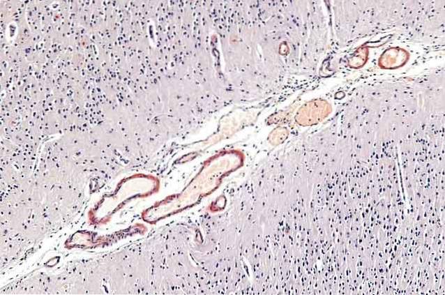
On the other hand, skin biopsy can be used for diagnosis. An immunostaining of skin samples taken from these patients can be a reliable test to detect the NOTCH3 protein, which is closely linked to the disease..
This technique can also show ultrastructural alterations in the blood vessels of the skin similar to those found in the cerebral arteries..
Cerebral microangiopathy progresses gradually throughout life and the level of affectation that it produces can be very heterogeneous, even within the same family.
The average age of onset of symptoms is 46 years. However, there are very isolated cases that have come to present symptoms at 8 years of age..
Generally, the prognosis is poor and most of those affected develop dementia and end up in bed needing constant care.
In fact, approximately 80% of those affected are in a situation of complete dependence somewhat before death. The life expectancy of these patients is not usually very long, with the average age of death being fixed at 68 years..
So far there is no definitive cure for cerebral microangiopathy, but treatments can be applied to combat the symptoms and make them change certain habits to improve the quality of life of the person, while preventing the progression of the disease.
As the Antioquia Neurosciences group indicates, it is important that these patients are properly diagnosed, since there are certain treatments that are not effective such as: triptans or drugs designed to combat migraine headaches, cerebral angiography or anticoagulant treatments.
In short, the use of drugs in this type of patient is not recommended because they may increase the risk of intracerebral hemorrhage or even produce no benefit..
However, there are some documented cases of the benefit of acetazolamide (ACZ) for the improvement of migraine typical of cerebral microangiopathy, but more research is needed.
The ideal is an interdisciplinary approach, combining:


Yet No Comments