
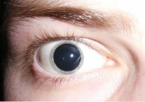
The mydriasis It is the increase in diameter or dilation of the central hole of the eye called the pupil. The pupil is the opening found in the colored portion of the eye, known as iris. The iris has two muscle groups that enlarge or decrease the size of the pupil in a reflected response to the amount of ambient light..
Thus, when the environment is illuminated, the pupil closes regulating the passage of light. On the contrary, if the environment is dark or with little light, the pupil dilates to allow the passage of as much light as possible and improve vision..
The decrease in the diameter of the pupil is called miosis while the increase in its diameter is known as mydriasis. Under normal conditions, both miosis and mydriasis occur simultaneously, but there may be variations caused by medications or pathological conditions..
In the case of the effect caused by the drugs, it is usually reversible once the treatment is stopped..
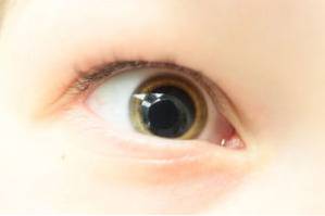
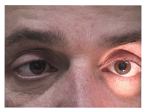
When asymmetry in the diameter of the pupils is observed, the patient must be studied deeply to discover the cause of this sign called anisocoria.
Article index
The eye is a spherical organ that has a transparent specialized tissue called cornea, which allows the entry of light rays. It has a posterior component that receives and processes light stimuli. This area is called retina.
Through complex neurological and physiological mechanisms, the eye allows the processing of stimuli and the clear vision of objects.
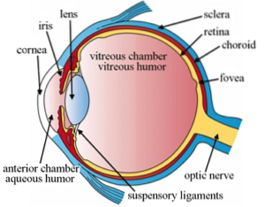
The colored portion of the eye is called iris. The iris is made up of two important muscle groups that vary the size of the central opening of the iris, called pupil.
The muscle group that is responsible for reducing pupil size is called sphincter muscle of the iris or sphincter pupil muscle, and the one in charge of increasing it is the iris dilator muscle or pupil dilator muscle.
The closing and opening of the pupil is a reflex mechanism that responds to light stimuli. This reflex regulates the amount of light that enters the eye.
In a well-lit environment, the sphincter muscle of the iris regulates the passage of light that the eye receives, reducing the pupil diameter. This process is known as miosis.
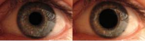
When the individual is in a dark environment, the dilator muscle of the pupil is responsible for expanding the size of the pupil to let in more light. The opening or increase in the diameter of the pupil is known as mydriasis.
The circumference of the pupil varies from 2 to 4 mm in response to bright light and from 4 to 8 mm in the dark. When there are no pathologies, this size is the same for both pupils..
Under normal conditions, mydriasis occurs as a response of the pupillary reflex. Whereas with exposure to light, the pupil contracts; with the absence of this, the pupil dilates.
Mydriasis is the normal response of the pupil dilator muscle to darkness. It occurs to let as much light pass through and be able to reproduce the image perceived through the eyes.
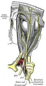
The reflex is carried out by the neurological response of a specialized component of the nerve common motorocular.
The final objective of these responses to light stimuli is to allow adequate vision of objects both in environments with bright or exaggerated lighting, as well as in those dark or with little light..
Both in natural conditions and in those caused by the doctor in the clinical evaluation, mydriasis is in both eyes equally. That is why normal mydriasis is said to be symmetric and bilateral.
When there is a problem or blockage in the neurological mechanism that regulates the activation of the dilator and constrictor muscles of the pupil, differences in the diameter of the pupils can be seen, a condition known as anisocoria, and, in severe cases, complete absence of response.
Abnormal mydriasis can be unilateral or bilateral and can be caused by pathophysiological, benign, or malignant causes, as well as pharmacological causes..
The common motor-ocular cranial nerve is a neurological component that has motor functions and reflex functions..
It is responsible for innervating several muscles that allow voluntary movement of the eye and through a specialized branch, it innervates the muscles that allow the variation of pupillary size..
Compression of this nerve by an external mass, whether tumorous, malignant or benign, or vascular, as in the case of aneurysms, causes variations in the normal response of the pupillary reflex.
For example, if there is a tumor that compresses the nerve of the right eye, that pupil will be unable to respond adequately to light stimuli, remaining open or in mydriasis, even when there is a significant light stimulus. In this case, the left pupil will have a normal response.
Also know as Adie's tonic pupil, is the most common cause of unilateral mydriasis caused by neurological degeneration.
Patients with this condition may be totally asymptomatic and, on occasion, mydriasis is discovered by a third party who notices the difference in the patient's pupillary size..
The syndrome is triggered by a viral or bacterial infection that causes neurological damage to the communication pathways of the iris muscles..
The pupil of these patients may have a slow response or be completely paralyzed to light stimuli.
Isolated and transient episodes of mydriasis are caused by overactivity of the neurological fibers that regulate the muscles of the iris.
The pathophysiological mechanism by which this response occurs is not fully understood. However, it is closely associated with conditions such as migraine, diabetes mellitus, and high blood pressure..
In these cases, the patient also has blurred vision, pain around the eye, and sensitivity to light..
Atropine eye drops are used to cause the pupil to dilate during the physical examination in the ophthalmology office..
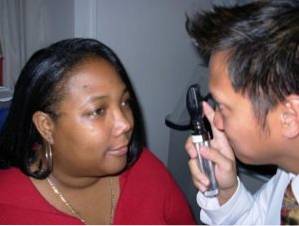
The direct effect of this type of topical medication is mydriasis. When the pupil is dilated, a proper evaluation of the retina can be made through the examination known as fundus.
Atropine drops are also used to treat some eye conditions.
Miller Fisher syndrome is a condition in which the body creates antibodies against its own nerves. This means that the body's protective system does not recognize nerve fibers and attacks them through special cells, causing their destruction as if they were a foreign organism..
The patient with this condition presents three typical symptoms that are, uncoordinated movements, decrease or absence of the reflex response and pupillary mydriasis with paralysis before the stimuli.
Although its cause is not exactly known, it is associated with viral infections such as chicken pox, rubella, cytomegalovirus and HIV, among others..
The aqueduct of Silvio is a brain structure that serves as a passage for the flow of cerebrospinal fluid.
When this brain area becomes blocked, it initiates a process of increasing the amount of intracranial fluid called hydrocephalus.
The increase in the amount of fluid inside the skull increases the intracranial pressure causing compression of the nucleus in which the nerves that regulate the movement of the pupil originate..
In these cases, dilated pupils are observed with little reaction to light stimuli..
When the physical examination of a patient with significant head trauma is performed, and it is found that his pupils are bilaterally dilated and there is no evidence of response to stimuli, this is considered a sign of irreversible brain damage.
The dilation response occurs by an increase in normal pressure within the skull due to brain swelling or blood pooling from trauma.
When mydriasis lasts more than 6 hours in this type of patient, it is an indicative factor of poor prognosis and most likely death of the injured person.
Some of the treatments for the control of psychiatric pathologies have an effect on the neurological signals that control pupillary movement..
Most of the time, the effect caused by these treatments is transitory and the response normalizes when they are stopped..
Tricyclic antidepressants, antipsychotics, and serotonin reuptake inhibitor drugs, as well as some migraine treatments, are drugs that are associated with bilateral mydriasis..
Illicit drugs with a stimulant effect such as amphetamines, cocaine and MDMA (ecstasy) cause significant dilation of the pupils, reaching a size that covers almost the entire iris.
Other drugs with hallucinogenic effect such as LSD (acid), hallucinogenic mushrooms and mescaline, are also causes of bilateral mydriasis.
In all these cases the pupils react to the light stimulus in a normal way and the mydriasis improves when the trigger factor is removed. However, other symptoms, such as eye movements and problems focusing vision, can persist and be irreversible..


Yet No Comments