The myosin it is a molecular motor, protein in nature, capable of moving on actin filaments in the cytosol. The energy that drives the displacement of myosin comes from the hydrolysis of ATP. Because of this, myosin is often defined as a mechanochemical enzyme.
In eukaryotes, myosin is a very abundant protein. There are different classes of myosin, which are encoded by a family of genes. In yeasts, 5 classes are distinguished, while in mammals dozens have been described.
Myosin has a wide variety of functions. Myosin I, together with actin, participates in the movement of keratocytes.
Myosin II provides rigidity to the plasma membrane, participates in cytokinesis and muscle contraction. Both myosins I and II collaborate with cell migration. Myosins I and V carry out vesicle transport along actin filaments.
Article index
In electron micrographs, the typical structure of myosin isoforms has three domains: head, neck, and tail. By hydrolysis with chymotrypsin, a segment consisting of the head and neck is obtained, called heavy meromyosin (HMM), and a segment of the tail, called light meromyosin (LMM).
The head domain is the N-terminal end of the heavy chain, and the tail domain is the C-terminal end of the light chain.
The classes of myosin can be differentiated by the number of polypeptide chains that compose it, and the abundance and class of light chain attached to the neck.
Myosin I has a polypeptide chain, which forms a head and its tail lacks alpha-helical regions. Whereas myosins I and V have two polypeptide chains, and therefore form two heads and a tail, in which the helical alpha chains coiled to form a rod-like structure.
Myosins I and V have binding sites for calmodulin, which regulates and binds Ca+two, on light chains. Myosin I fixes Ca+two in light chains, but does it differently than calmodulin.
At the mechanochemical level, myosins have three characteristics, namely:
- The myosin head is the motor domain that advances in discrete steps: The union of the myosin head to an actin filament, its inclination and subsequent separation produce the movement of myosin. This process is cyclical and depends on ATP.
- Conformal changes: the hydrolysis of an ATP molecule is coupled to each step of a myosin molecule, through levels of amplification and transmission. This involves large conformational changes of myosin..
The first level of amplification is produced by the loss of the gamma-phosphate group of ATP, which allows a reorganization of the structural elements in the ATP binding site. This rearrangement is coordinated with structural changes in the actin-binding site..
The second level of amplification involves the communication of the conformational change in the active site to structural components of the carboxyl terminal..
- Directionality: Myosins have been found to have polarity, or reverse directionality, toward the (+) end of the actin filament. This conclusion comes from the actin filament slippage experiments, using a fluorescence light microscope..
Myosin, together with actin, participates in muscle contraction, cell adhesion, cytokinesis, the stiffening of cortical membranes and the displacement of some vesicles, among other functions..
Defects in myosin can produce pathological conditions. For example, defects in myosins I and V are related, respectively, to myosin myopathies and pigmentation disorders (Griscelli syndrome). While disorders in myosin VI isoforms cause hearing loss.
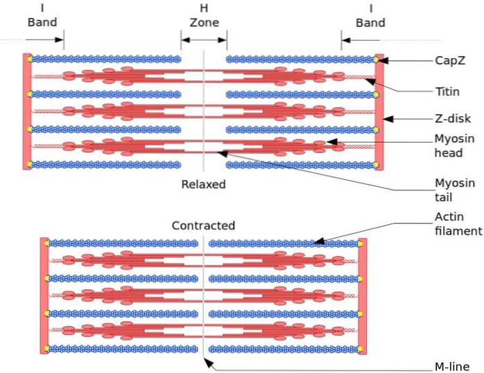
The functional and structural unit of skeletal muscle is the sarcomere. During muscle contraction, the length of the sarcomere reaches 30% of its original length.
Sarcomeres are made up of thick myosin filaments and thin actin filaments that are organized in a complex way. In general, the myosin heads are located at the distal ends of the filament and their tails towards the center of the sarcomere, and the organization is bipolar.
For muscle contraction to occur, the myosin heads at opposite ends must move toward the Z disk or the (+) end of the filament. Because the organization of the thick filaments is bipolar, the sliding of the thin filaments on the thick filaments occurs, driven by ATP.
The displacement force occurs because hundreds of myosin heads, of a thick filament, interact with a thin filament.
During mitosis, when microtubules at the spindle poles separate, actin and myosin II form a contractile ring at the equator of the cell. This ring contracts, decreasing its diameter and dividing the cell into two parts..
In mutant cells lacking myosin II, the plasma membrane easily deforms when an external force is applied. This happens because myosin II provides aggregation force to plasma membrane proteins..
In epithelial tissue, the contractile bundles of actin and myosin II are located in the vicinity of the plasma membrane, and form a circular girdle that surrounds the inner cell surface. This circular girdle determines the shape of the cell and maintains the bond between cells..
The contact between cells occurs by the union of the circular girdle to the cellular adhesion molecules, by means of union proteins.
Experimental evidence reveals that myosin V performs membrane transport from the Golgi apparatus to the periphery of the cell. Some evidences are:
- In nervous tissue cells, by astrocyte immunofluorescence it was found that myosin V is located next to the Golgi.
- In yeast, mutations in the myosin V gene disrupt protein secretion and consequently protein accumulates in the cytosol.
- The isoforms of myosin I are responsible for the transport of vacuoles towards the cell membrane. Using specific antibodies against myosin I isoforms, it was found that these isoforms are located in different parts of the cell..
For example, when a living amoeba is labeled with an antibody against myosin IC, the transport of the vacuole to the membrane is stopped. Because of this, the vacuole expands and the cell bursts..
There are numerous genes and mutations that cause hearing loss. This disease is frequently monogenetic .
Unconventional myosin mutations, with one or two myosin heads, affect the functioning of the inner ear. Some of the mutated myosin isoforms are myosin IIIA, myosin VIIA, and myosin XVA. Recently, two mutations were discovered in myosin VI.
Mutations in myosin VI are c.897G> T and p.926Q. The first mutation affects a region that interacts with the active site, called Switch I. Homozygous for the mutation exhibit the phenotype early, causing severe effects.
The second mutation affects a region of charged residues, in an alpha helix in the tail of myosin VI. This region is important for proximal motor dimerization, and affects the stereo-ciliary function of myosin VI..
Another mutation is p.Asn207Ser, which produces a motor incapable of producing force. This is because Asn 207 is an amino acid residue of the active site, whose function is the binding and hydrolysis of ATP.
The p.Arg657Trp mutation causes loss of myosin VI function. The Arg residue is involved in the conformational changes that couple hydrolysis to the movement of myosin.
Myosin X (Myo10) is an unconventional myosin that is expressed in the brain, endothelium, and many epithelia. Myo10 and three classes of actin-based projections (filopodia, invadopodia, and filopodia-like projections) work during cancer metastasis.
Invasive cancer cells have large numbers of filopodia and express high levels of fascina. This protein makes crosslinks between actin filaments. In order to escape the primary tumor, invadopodia are formed, rich in proteolytic activity, which digest the surrounding extracellular matrix..
Once the cells reach the extracellular matrix, the filopodia-like projections help to disperse and colonize. High levels of Myo10 indicate high aggressiveness and metastasis in breast cancer.
MyoX silencing causes loss of metastatic character of cells, which are unable to form actin-based projections. All these projections have integrin-based adhesions, which are carried by Myo10 within the filopodium..
MyoX is involved in the formation of the centrosome. The absence of MyoX favors the formation of multipolar spindles. MyoX is also involved in signaling in cancer cells. For example, MyoX is activated by 3,4,5, -inositol triphosphate (PIP3).


Yet No Comments