
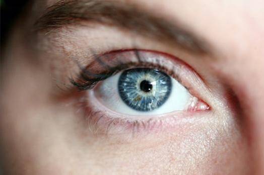
The miosis It is the contraction of the pupil of the eye. This is a normal response that limits the amount of light that enters the eyeball under bright lighting conditions. It is the final result of the photomotor reflex, which is responsible for the contraction of the pupil (miosis) when there is a lot of light in the environment, being the normal the pupillary contraction of both eyes in response to the lighting conditions..
However, not in all cases miosis is normal, in fact when it occurs in low lighting conditions, it is accompanied by other symptoms (such as drowsiness or disorientation). When it occurs in only one eye, it should be considered pathological.
It is extremely important to determine the cause since it is usually due to serious conditions that can compromise the life of the person.
The evaluation of the miosis is very simple, it is enough to observe directly the eye of the person and determine the diameter of the pupil; as long as it is 2 mm or less, it will speak of miosis.
Article index
Miosis is in most cases a normal response to external lighting conditions and represents the visible clinical sign of the activation of the photomotor reflex..
When said reflex is altered, either by organic lesions or as a consequence of the effect of toxic substances or medications, it is said that it is a pathological miosis, being necessary a complete physical examination to determine the cause and correct it..
In order to understand miosis well, it is essential to know its mechanism (physiology); once this is done, it will be easier to identify the different pathologies that trigger a pathological miosis.
The photomotor reflex begins when light enters the eyeball and stimulates the photoreceptor cells located in the retina (cones, rods, photoreptor ganglion cells), converting the light into an electrical impulse that travels through the sensory fibers of the second to cranial (ophthalmic nerve) to the midbrain.
In this region the impulse reaches the pretectal nucleus located in the superior colliculus, this without passing through the lateral geniculate nucleus or the visual cortex, therefore the reflex is exclusively integrated in the midbrain without the participation of superior structures.
Once the sensory impulse reaches the pretectal nucleus, it stimulates the neurons that link it with the visceromotor nucleus of Edinger-Westphal, from where parasympathetic motor fibers that accompany the third cranial nerve (oculomotor nerve) start..
Once the third cranial nerve enters the orbit, the accompanying parasympathetic fibers enter the ciliary ganglion from where postganglionic motor fibers known as short ciliary nerves exit, which will ultimately be responsible for the contraction of the ciliary muscle in response. to the light.
It is known as direct photomotor reflex to the contraction of the pupil (miosis) in response to the direct stimulus of light on the same eye; that is, light enters the right eye and the right pupil contracts.
In addition to the direct photomotor reflex, there is what is known as the consensual reflex, which consists of contralateral pupil contraction in response to the light stimulus in the opposite eye; for example, light stimulates the right eye and the pupil of the left eye contracts.
The consensual reflex is responsible for both pupils having the same degree of miosis, therefore it is expected that under normal conditions the pupils are symmetrical. When this does not occur, a damage to the reflex integration path should be considered..
When miosis occurs in low-light conditions, is asymmetric (one eye is yes and the other is not) or is accompanied by other clinical symptoms such as confusion, disorientation or altered state of consciousness, a pathological miosis should be considered.
The causes of pathological myosis are multiple and very varied, being the subject of extensive medical treatises, however, from the general point of view, two large groups of causes can be considered:
- Lesions of the pathway of integration of the photomotor reflex.
- Effects of toxic substances, medications or drugs.
In general, the clinical history of the patient, the findings of the physical examination and the complementary examinations (tomography, toxicological tests or other as the case may be), allow to establish with precision the cause of the pathological myiosis, this being of vital importance since According to the cause, the treatment must be decided.
The photomotor and consensual reflex chain can be affected at various points, from lesions in the retina that prevent the light stimulus from becoming an electrical stimulus, to alterations in the motor nerves that prevent the contraction of the ciliary muscle in response to light.
There are countless pathologies and lesions that can alter the photomotor reflex inducing pathological miosis, the most frequent being some types of cerebral hemorrhages (such as pontine hemorrhages), Horner syndrome, Pancoast tumor and cluster headache, to mention only some of the most common causes.
In Horner syndrome, there is involvement of the sympathetic fibers responsible for mydriasis (dilation of the pupil), which is why the balance between myiosis and mydriasis is lost in response to different ambient light conditions..
When this occurs, the neurovegetative innervation of the eye is commanded exclusively by the parasympathetic system, which, since there is no one to antagonize it, produces a sustained and pathological miosis of the eye whose sympathetic pathway is compromised..
A rare but very serious cause of miosis is Pancoast tumor, a type of lung cancer that involves the apex of the organ by infiltrating adjacent structures including the cervical sympathetic nodes. When this occurs there is involvement of the sympathetic fibers, as occurs in Horner syndrome.
On the other hand, in cluster headache there is transitory abolition of mydriasis due to a not yet well defined pathological alteration of the sympathetic pathway, leaving once again the neurovegetative innervation commanded by the parasympathetic, which induces sustained miosis as it lacks natural antagonism. sympathetic system.
The medications, drugs and toxins that can exert effects on the parasympathetic system are many and of various types, however there is a common denominator that allows us to suspect the toxic effects of some substance as responsible for the miosis: the associated neurological symptoms.
Neurological signs such as stupor, confusion, drowsiness, agitation, sensory disturbance, or motor disability will generally present in any patient with drug- or drug-induced miosis..
It all depends on the type of substance involved in miosis, this being the most obvious difference with respect to organic lesions, however the possibility of brain hemorrhages should never be ignored, which can sometimes be very similar to poisonings.
Substances that cause miosis include:
- All opioid derivatives
- Cholinergic agents (such as acetylcholine)
- Acetyl cholinesterase inhibitors (neostigmine, physostigmine)
- Nicotine
- Parasympathomimetics (such as pilocarpine, a medicine commonly used to treat glaucoma)
- Antipsychotic drugs (such as haldol and risperidone)
- Some antihistamines like diphenhydramine
- Imidazolines, including the antihypertensive clonidine
The treatment of miosis will depend largely on the cause, in fact physiological miosis does not require any treatment, as well as that which occurs as a side effect of a drug used to treat a known pathology (pilocarpine, clonidine, etc.).
In those cases where treatment is required, it will generally be necessary to identify the cause and initiate appropriate treatment for the specific cause, provided that one is available; This implies that the miosis itself is not treated since it constitutes a symptom, so the underlying disease responsible for it must be attacked.

Yet No Comments