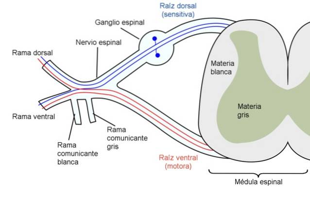
A myotome It is a set of muscle fibers innervated by a segmental, spinal or spinal root or nerve. The motor axons of each segmental root or nerve innervate several muscles, and almost all muscles are innervated by more than one segmental nerve and thus by an equivalent number of spinal segments..
In vertebrates, the dermatomes of the skin, the myotomes of skeletal muscle, and the sclerotomes of the vertebrae have a common embryological origin, the somites. These derive from the mesoderm and develop on each side and along the neural tube..
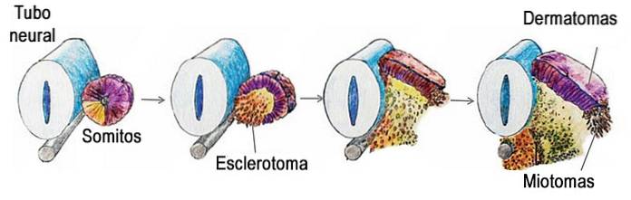
demarcation of the myotomes was relatively easier than that of the other segments derived from somites, such as sclerotomes and dermatomes.
This is due to the fact that the injury to a segmental root or nerve causes the immediate loss of function of the skeletal muscle innervated by said nerve and, consequently, the loss of the corresponding motility, which is easily detected in the neurological examination..
Muscle weakness, paralysis or absence of contraction and alteration of the tendon reflexes are signs that allow evaluating the different myotomes of the musculoskeletal system.
Article index
The evaluation of myotomes is generally used by some clinicians, by neurologists, traumatologists and, above all, by physical therapists.
In the neurological examination, detailed tests of each myotome allow to evaluate the integrity of the motor system related to each of the examined myotomes. These tests examine isometric contractions under resistance and tendon reflexes..
The absence of any of the functions examined and corresponding to a particular myotome allows the lesion to be located in the medullary segment or in the root or segmental nerve corresponding to the myotome examined.
On some occasions, when examining a certain myotome, there is not a total loss of function, but rather a weakness in the muscle contraction of the muscle group or groups corresponding to the myotome examined.
In these cases, the lesion can be located in the segmental nerve and one of the most frequent causes is root compression due to herniation of the intervertebral disc. The affected myotome allows to locate the intervertebral disc that is compressing the root.
The roots related to the muscular functions of myotomes corresponding to the upper and lower extremities are shown below..
Spinal Root C1 and C2 → Muscles that flex and extend the neck
Spinal root C3 → Muscles that flex the neck laterally
Spinal Root C4 → Muscles that raise the Shoulder
Spinal root C5 → Muscles that produce shoulder abduction
Spinal Root C6 → Elbow Flexor and Wrist Extensor Muscles
C7 spinal root → Elbow extensors and wrist flexors
Spinal root C8 → Extensor muscles of the fingers of the hand
Spinal root T1 → Muscles that produce thumb abduction
Spinal root L2 → Muscles that flex the hip
Spinal root L3 → Muscles that produce knee extension
Spinal root L4 → Muscles responsible for dorsiflexion of the ankle
Spinal root L5 → Extensor muscles of the toes
Spinal root S1 → Muscles that produce plantar flexion of the ankle
Spinal root S5 → Knee flexor muscles
When examining muscle functions, the examiner exerts resistance against the action of the corresponding muscle. For example, for the right lateral flexion of the head, the examiner exerts force against this movement and in this way the myotome corresponding to the C3 root is evaluated.
To describe the anatomical distribution of myotomes, although there are many variants, the distribution of the peripheral nerves, the medullary root of origin, as well as the related muscles are discussed below. Tendon reflexes and related roots are also included..
Axillary → C5 and C6
Supraclavicular → C3 and C4
Suprascapular → C5 and C6
Thoracic (long) → C5, C6 and C7
Musculocutaneous → C5, C6 and C7
Medial cutaneous forearm → C8 and T1
Lateral cutaneous forearm → C5 and C6
Posterior cutaneous of the forearm → C5, C6, C7 and C8
Radial → C5, C6, C7, C8 and T1
Medium → C6, C7, C8 and T1
Ulnar → C8 and T1
Pudendo → S2, S3 and S4
Lateral cutaneous thigh → L2 and L3
Medial cutaneous thigh → L2 and L3
Intermediate cutaneous thigh → L2 and L3
Posterior cutaneous of the thigh → S1, S2 and S3
Femoral → L2, L3 and L4
Shutter → L2, L3 and L4
Sciatic → L4, L5, S1, S2 and S3
Tibial → L4, L5, S1, S2 and S3
Common peroneal → L4, L5, S1 and S2
Superficial peroneum → L4, L5 and S1
Deep peroneal → L4, L5, S1 and S2
Lateral cutaneous leg → L4, L5, S1 and S2
Saphene → L3 and L4
Sural → S1 and S2
Medial plantar → L4 and L5
Plantar Lateral → S1 and S2
Each nerve root and its corresponding muscle are listed below:
C2 → Longus Colli, sternocleidomastoid and rectum capitis
C3 → Trapezius and splenius capitis
C4 → Trapezius and levator scapulae
C5 → Supraspinatus, infraspinatus, deltoid and biceps
C6 → Biceps, supinator, wrist extensors
C7 → Triceps and wrist flexors
C8 → Ulnar deviator, extensor pollicis, and adductor pollicis
L2 → Psoas, adductor hip
L3 → Psoas and quadriceps
L4 → Tibialis anterior, extensor hallucis
L5 → Extensor hallucis, fibulae, gluteus medius and ankle dorsiflexors
S1 → Buttocks, peroneals and plantar flexors
S2 → Glutes and plantar flexors
S4 → Bladder and recti
Myotomes or segmental innervation of skeletal muscles are related to osteotendinous reflexes and their assessment allows evaluating the integrity of the motor and sensory pathways, as well as the corresponding spinal segments..
- Achilles reflex → S1 and S2
- Patellar Reflex → L2, L3 and L4
- Lower cutaneous-abdominal → T10-T12
- Middle abdominal-cutaneous → T8 and T9
- Upper cutaneous-abdominal → T6 and T7
Bicipital Reflex → C5, C6
Tricipital Reflex → C6, C7, C8
Radial Reflex → C5, C6 and C7
Some integrated examples including the root, muscles, function, and innervation of various myotomes are:
C5 → Biceps → Elbow flexion → Bicipital → Musculoskeletal
C7 → Triceps Brachii → Elbow Extension → Tricipital → Radial
L3 → Quadriceps crural → Knee extension → Patellar → Femoral

Yet No Comments