Mitochondria are characteristic intracellular organelles of all eukaryotic cells. They are in charge of an important part of cellular energy metabolism and are the main site of ATP production in cells with aerobic metabolism..
Seen under the microscope, these organelles are similar in size to that of a bacterium and share many of their genetic characteristics with prokaryotes, such as the presence of a circular genome, bacterial ribosomes, and transfer RNAs similar to those of other prokaryotes..

Endosymbiotic theory proposes that these organelles arose in eukaryotic progenitors millions of years ago from prokaryotic cells that "parasitized" primitive eukaryotes, giving them the ability to live in aerobiosis and to use oxygen for energy, receiving shelter in return. and nutrients.
Since their genome must have been reduced, the formation of these organelles became dependent, to a great extent, on the importation of proteins that are synthesized in the cytosol from genes encoded in the nucleus, also from phospholipids and other metabolites, for which were adapted complex transport machinery.
Today it is known that the mitochondria function as the "sources of power" of all aerobic eukaryotic cells and that the Krebs cycle occurs in them, the synthesis of pyrimidines, amino acids and some phospholipids. Inside it, the oxidation of fatty acids also occurs, from which large amounts of ATP are obtained..
As in all cellular organisms, mitochondrial DNA is prone to mutations, resulting in mitochondrial dysfunctions that lead to neurodegenerative disorders, cardiomyopathies, metabolic syndromes, cancer, deafness, blindness, and other pathologies.
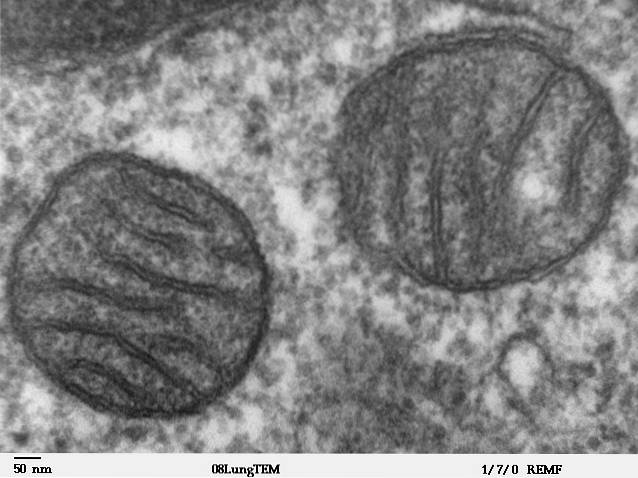
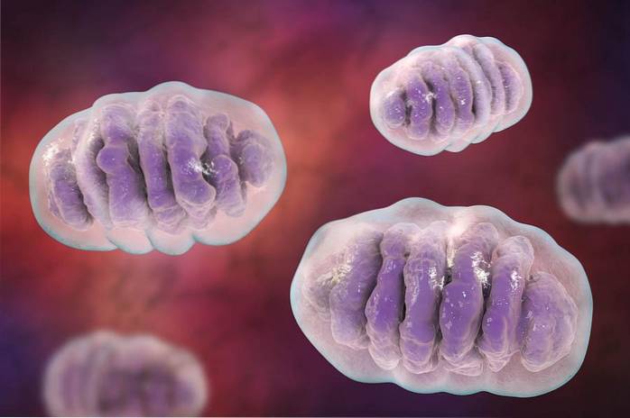
Mitochondria are quite large cytosolic organelles, their size exceeds that of the nucleus, vacuoles and chloroplasts of many cells; its volume can represent up to 25% of the total volume of the cell. They have a characteristic worm- or sausage-like shape and can measure several micrometers in length..
They are organelles surrounded by a double membrane that have their own genome, that is, inside there is a DNA molecule foreign (different) to the DNA contained within the cell nucleus. They also have ribosomal RNA and transfer RNA of their own..
Despite the above, they depend on nuclear genes for the production of most of their proteins, which are specifically marked during their translation in the cytosol to be transported to the mitochondria..
Mitochondria divide and multiply independently of cells; their division occurs by mitosis, which results in the formation of a more or less exact copy of each one. In other words, when these organelles divide they do so by "splitting in half".
The number of mitochondria in eukaryotic cells is highly dependent on the type of cell and its function; that is, in the same tissue of a multicellular organism, some cells may have a greater number of mitochondria than others. An example of this are cardiac muscle cells, which have an abundant number of mitochondria.
Mitochondria are essential organelles for aerobic cells. These function in the integration of intermediate metabolism in several metabolic pathways, among which oxidative phosphorylation for the production of ATP in cells stands out..
Inside it occurs the oxidation of fatty acids, the Krebs cycle or tricarboxylic acids, the urea cycle, ketogenesis and gluconeogenesis. Mitochondria also play a role in the synthesis of pyrimidines and some phospholipids.
They are also involved in part in the metabolism of amino acids and lipids, in the synthesis of the heme group, in calcium homeostasis and in the processes of programmed cell death or apoptosis.
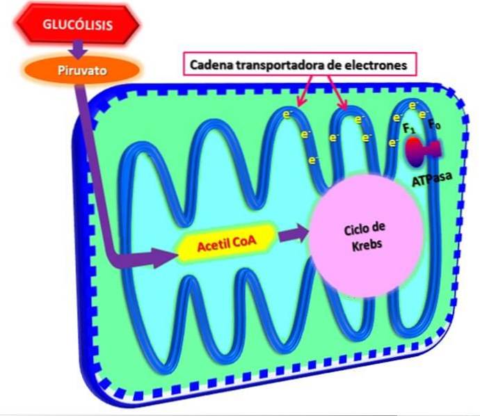
Glycolysis, the process of oxidizing glucose to extract energy from it in the form of ATP, occurs in the cytosolic compartment. In cells with aerobic metabolism, pyruvate (the end product of the glycolytic pathway per se) is transported to the mitochondria, where it serves as a substrate for the pyruvate dehydrogenase enzyme complex.
This complex is responsible for the decarboxylation of pyruvate to CO₂, NADH and acetyl-CoA. It is said that the energy of this process is "stored" in the form of acetyl-CoA molecules, since these are the ones that "enter" the Krebs cycle, where their acetyl portion is completely oxidized to CO₂ and water..
In the same way, the lipids that circulate through the bloodstream and enter the cells are oxidized directly in the mitochondria through a process that begins at the carbonyl end of them and by which two carbon atoms are eliminated simultaneously in each " return", forming one acetyl-CoA molecule at a time.
The degradation of fatty acids ends with the production of NADH and FADH2, which are molecules with high-energy electrons that participate in oxidation-reduction reactions..
During the Krebs cycle, CO₂ is eliminated as a waste product, meanwhile the NADH and FADH2 molecules are transported to the electron transport chain in the inner membrane of the mitochondria, where they are used in the oxidative phosphorylation process..
Enzymes that participate in the electron transport chain and oxidative phosphorylation are found in the inner membrane of the mitochondria. In this process, the NADH and FADH2 molecules serve as “transporters” of electrons, as they pass them from the oxidizing molecules to the transport chain..
These electrons release energy as they pass through the transport chain and this energy is used to eject protons (H +) from the matrix into the intermembrane space through the inner membrane, generating a proton gradient..
This gradient functions as an energy source that is connected to other reactions that require energy, such as, for example, the generation of ATP by phosphorylation of ADP..
These organelles are unique among other cytosolic organelles for several reasons, which can be understood from knowledge of their parts..
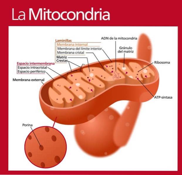
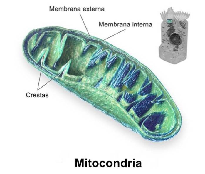
Mitochondria, as already mentioned, are cytosolic organelles surrounded by a double membrane. This membrane is divided into the outer mitochondrial membrane and the inner mitochondrial membrane, very different from each other and separated from each other by the intermembrane space..
This membrane is the one that serves as the interface between the cytosol and the mitochondrial lumen. Like all biological membranes, the outer mitochondrial membrane is a lipid bilayer to which peripheral and integral proteins are associated.
Many authors agree that the ratio between proteins and lipids in this membrane is close to 50:50 and that this membrane is very similar to that of Gram negative bacteria.
The proteins of the outer membrane function in the transport of different types of molecules towards the intermembrane space, many of these proteins are known as "porins", since they form channels or pores that allow the free passage of small molecules from one side to the other. other.
This membrane contains a very large number of proteins (almost 80%), much greater than that of the outer membrane and one of the highest percentages in the whole cell (the highest protein: lipid ratio).
It is a membrane less permeable to the passage of molecules and forms multiple folds or ridges that project towards the lumen or the mitochondrial matrix, although the number and arrangement of these folds varies considerably from one type of cell to another, even in the same organism.
The inner mitochondrial membrane is the main functional compartment of these organelles and this is essentially due to their associated proteins.
Its folds or ridges play a special role in increasing the membrane surface, which reasonably contributes to the increase in the number of proteins and enzymes that participate in mitochondrial functions, that is, in oxidative phosphorylation, mainly (electron transport chain)..
As can be inferred from its name, the intermembrane space is the one that separates the outer and inner mitochondrial membranes.
Since the outer mitochondrial membrane has many pores and channels that facilitate the free diffusion of molecules from one side of it to the other, the intermembrane space has a composition quite similar to that of the cytosol, at least with respect to ions and certain molecules. small in size.
The mitochondrial matrix is the internal space of the mitochondria and is the place where the mitochondrial genomic DNA is found. In addition, in this "liquid" there are also some of the important enzymes that participate in cellular energy metabolism (the amount of proteins is greater than 50%).
In the mitochondrial matrix there are, for example, the enzymes belonging to the Krebs cycle or the tricarboxylic acid cycle, which is one of the main routes of oxidative metabolism in aerobic organisms or cells..
Mitochondria are unique cytosolic organelles in cells, since they have their own genome, that is, they have their own genetic system, which is different from that of the cell (enclosed in the nucleus).
The genome of mitochondria consists of circular DNA molecules (such as that of prokaryotes), of which there may be several copies per mitochondrion. The size of each genome depends a lot on the species considered, but in humans, for example, this is about 16 kb.
The genes that code for some mitochondrial proteins are found in these DNA molecules. There are also the genes that code for ribosomal RNAs and transfer RNAs that are necessary for the translation of the proteins encoded by the mitochondrial genome inside these organelles..
The genetic code used by mitochondria to "read" and "translate" the proteins that are encoded in their genome is somewhat different from the universal genetic code.
Human mitochondrial diseases are a fairly heterogeneous group of diseases, since they have to do with mutations in both mitochondrial and nuclear DNA.
Depending on the type of mutation or genetic defect, there are different pathological manifestations related to the mitochondria, which can affect any organ system in the body and people of any age.
These mitochondrial defects can be transmitted from one generation to another through the maternal route, through the X chromosome, or through the autosomal route. For this reason, mitochondrial disorders are truly heterogeneous both in the clinical aspect and in the tissue-specific manifestations..
Among some of the clinical manifestations related to mitochondrial defects are:
Animal cells and plant cells contain mitochondria. In both types of cells these organelles perform equivalent functions and, although they are not very important, there are some small differences between these organelles.
The main differences between animal and plant mitochondria have to do with morphology, size, and some genomic characteristics. Thus, mitochondria can vary in size, number, shape and organization of the inner ridges; although this is also true for the different types of cells in the same organism.
The size of the mitochondrial genome of animals is slightly smaller than that of plants (̴ 20kb vs 200kb, respectively). Furthermore, unlike animal mitochondria, those in plant cells encode three types of ribosomal RNA (animals encode only two).
However, plant mitochondria depend on some nuclear transfer RNA for the synthesis of their proteins.
In addition to those already mentioned, there are not many other differences between the mitochondria of animal cells and plant cells, as reported by Cowdry in 1917..

Yet No Comments