
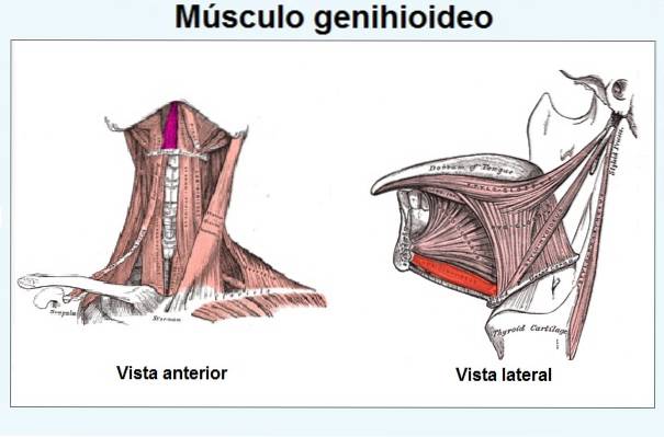
The geniohyoid muscle, together with the digastric, stylohyoid and mylohyoid muscles they make up the muscle group called suprahyoid. The muscle originates from the chin of the jaw and goes to the hyoid bone where it inserts. Its name comes from the Latin musculus geniohyoideus.
This muscle, together with the rest of the members of the suprahyoid group, forms the floor of the mouth and participates in the movement of the hyoid bone forward and upward during swallowing. It also helps in the movement of the jaw down.
The geniohyoid is a small, cylindrical, deep and even muscle. Both muscles (right and left) are finely united in the midline of the hyoid bone by a poorly differentiated simple tissue. Occasionally, this junction can become so strong that it simulates being a single, centrally located, odd muscle..
The genioglossus muscle is related as follows: below it is the mylohyoid muscle and above it it is related to the genioglossus muscle, as well as with the mucosa of the floor of the mouth and with the sublingual gland.
On the other hand, the ventral branch of the C1 spinal nerve is in charge of innervating the geniohyoid muscle. This runs accompanied by the twelfth cranial nerve (hypoglossal nerve), and is supplied by the lingual and sublingual arteries.
The geniohyoid muscle is not exempt from hypertension or being affected by trigger points. This condition causes very annoying symptoms such as headaches, difficulty swallowing, pain in the neck, sore throat, among others. Finally, very sporadic cases have been reported of the presence of an accessory aberrant fascicle that arises from it and inserts into the hyoid bone in its greater horn..
Article index
It originates as thin and short tendons that arise in the lower mental spine or in the lower geni processes, as this anatomical site was previously known.
From the site of origin, the muscle runs backward and downward, until it reaches the medial area of the anterior aspect of the hyoid bone where it is inserted. During the journey the tendinous fibers thicken to form the body of the muscle.
The fibers of the spinal nerve C1 penetrate the geniohyoid muscle from the deepest or internal zone of this to innervate it and their fibers run along the hypoglossal nerve (cranial nerve XII).
The supply of the geniohyoid muscle is provided by a collateral extension of the external carotid, called the lingual artery. From the latter originates the sublingual artery that also supplies the geniohyoid muscle.
The geniohyoid is one of the muscles of the neck that supports the hyoid bone, which is the only bone that is suspended and supported only by muscles, since it does not articulate with any other bone..
In this sense, the neck muscles, including the geniohyoid, interconnect the hyoid bone with the head. These four muscles perform their functions in pairs with their respective counterparts..
On the other hand, the functions of the geniohyoid muscle will depend on the point of support that the muscle adopts. If it rests on the hyoid bone when it is contracted and immobilized, it lowers the jaw and pulls it back, shortening the floor of the mouth and widening the pharynx, that is, it acts when the mouth is opened.
If, on the contrary, it rests on the jaw, then it is able to raise the hyoid bone, at the same time as it moves it forward. That is why, it is said that it is an antagonist of the stylohyoid and masseter muscles, who do the opposite.
These movements occur during swallowing. This muscle also helps in sucking and in the movement of the tongue in an anterior direction..
It should be noted that the descent of the jaw is not the only function it exerts on it, since the suprahyoid group controls the dynamics of the levator and propulsion muscles of the jaw.
On the other hand, the four suprahyoid muscles need the proper functioning (contraction) of the infrahyoids to work correctly, since the good performance of the geniohyoid muscle and the anterior muscles of the neck in general depends on the existence of a balance in the postural position. orthostatic hyoid bone.
Cervical headaches are a very common affection and many of them are related to myofascial problems at the level of the neck muscles. That is, the presence of trigger points or sore points.
In pain therapy sessions the aim is first to eliminate the trigger point and then to stretch and relax the muscles involved. Trigger points can be found at the neck level, although the geniohyoid is not the most vulnerable, in these cases the omohyoid muscle is more affected.
However, its involvement is not ruled out, since the geniohyoid muscle can become stressed (muscular hypertonia) due to abnormal functioning of the first vertebra (atlas) or as a consequence of strong emotional reactions..
The tension and appearance of trigger points in any of the deep muscles of the neck, including the geniohyoid, can cause the following symptoms: sore throat, difficulty in swallowing food, sensation of pain when speaking, pain in the neck, headaches, pain lingual, among others.
A study carried out by Carulla et al. In 2008 determined the influence of mouth or nasal breathing on the position of the hyoid bone..
The authors found certain differences between the two groups. In the group of mouth breathers, they observed that the mylohyoid, geniohyoid and anterior belly of the digastric muscles were more elongated compared to the control group.
This occurs due to the greater resistance exerted by the median constrictor muscles of the pharynx, stylohyoid, posterior digastric belly and stylohyoid ligament to the anterior transfer of the hyoid bone; movement performed by the mylohyoid, geniohyoid, and anterior digastric belly muscles during mouth breathing.

Yet No Comments