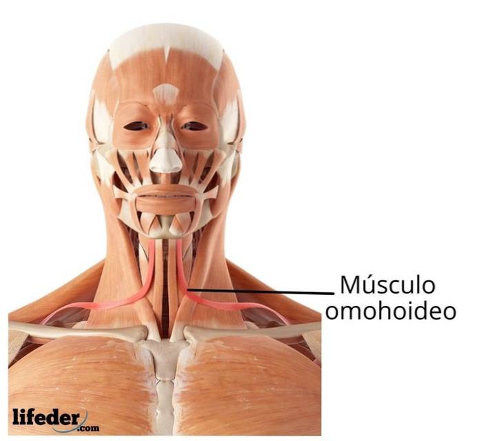
The omohyoid muscle It is a long, flattened, thin neck muscle. Morphologically it is characterized by being one of the few digastric muscles of the human body, its bellies are consecutive and are joined by an intermediate tendon.
This bilateral muscle is also called the omoplatohyoid or scapulohyoid, due to its bony attachments to the scapula or shoulder blade, and the hyoid bone. The path of this muscle is upwards and towards the center. It belongs to the anterior group of neck muscles, within the infrahyoid subclassification.
This means that its origin and insertion is below the hyoid bone. The infrahyoid muscles are classified as superficial and deep; the omohyoid muscle is located within the superficial muscles, being the most superficial of this group and, at the same time, the most lateral. Be part of those responsible for swallowing and phonation.

To describe the origin and insertion of the omohyoid muscle, the anatomy of the scapula must be broadly remembered. The scapula or shoulder blade is an even, median and triangular bone that is located in the posterolateral region of the thorax. In this two faces, three edges and four angles are described.
The omohyoid muscle originates from the upper edge of the scapula. The main feature of the upper border is the scapular or coracoid notch.
This notch is converted into a foramen by the presence of a ligament: the transverse scapular ligament or coracoid ligament. This crosses it transversely at the upper end; the suprascapular nerve passes through this foramen.
The omohyoid muscle originates from the transverse scapular ligament within the scapular or coracoid notch and some fibers insert on the superior edge of the scapula proper, medial to the notch.
From there, it travels forward, up, and toward the center, passing over the vascular axis of the neck and posterior to the sternocleidomastoid muscle..
During its course, it forms in its middle part a tendon called the intermediate tendon of the omohyoid muscle, which gives it the characteristic of a digastric muscle. It has a lower and an upper belly, or a posterior and an anterior belly due to its route, which becomes ventral as it rises.
It continues with the upper or anterior belly, which has an almost completely vertical upward direction, attaches to the lower border and the greater horn of the hyoid bone, laterally to the sternohyoid muscle.
In the lower abdomen, on its anterior face, it is related to the trapezius muscle, the clavicle and the subclavian muscle.
As it ascends, it becomes more superficial, and relates only to the deep cervical fascia and the skin. This deep cervical fascia wraps it at the level of the intermediate tendon and fixes it.
The upper belly, also on its anterior face, is related to the sternocleidomastoid muscle, and when inserting into the hyoid it leaves the shadow of the sternocleidomastoid and becomes superficial again.
The lower belly of the omohyoid is related by its posterior aspect with the serratus major muscle, ascends and is related to the brachial plexus, the scalene muscles and the neurovascular bundle of the neck.
The intermediate tendon is located on the jugular vein; This is why the tendon is sometimes used to identify the internal jugular vein in neck dissections..
The upper belly, almost vertical, is related to the sternothyroid and thyrohyoid muscles, which separate the omohyoid muscle from the thyroid gland..
The omohyoid muscle is part of the structures that delimit the carotid triangle, one of the most important triangles in the anatomy due to its content and because it represents a part of the anterior cervical triangle.
The carotid triangle is made up of the anterior border of the sternocleidomastoid muscle in the posterior part, the posterior belly of the digastric muscle anterosuperiormente, and the superior belly of the omohyoid muscle anteroinferiormente.
In this triangle is located the carotid bifurcation (hence its name), the internal jugular vein, the hypoglossal nerve, the cervical loop of the cervical plexus and the vagus nerve, as well as the internal branch of the superior laryngeal nerve..
The main function of the omohyoid muscle is to depress and fix the hyoid bone as well as the larynx; this is done to facilitate swallowing and phonation.
It is also responsible for tightening the cervical fascia to ensure the patency of the internal jugular vein.
Omohyoid muscle syndrome is called a rare-onset pathology, the main characteristic of which is the appearance of a lateral mass in the neck when swallowing due to dysfunction of the omohyoid muscle..
Studies indicate that this dysfunction is mainly due to the union of the cervical fascia with the intermediate tendon giving way or stretching..
The problems caused in this pathology are mainly aesthetic, as well as the anxiety of the patient when viewing the lateral mass since he fears it may be caused by some tumor pathology.
The omohyoid muscle receives its blood supply through branches of the inferior thyroid artery, which arises from the subclavian artery.
From there, the esophagus, larynx, trachea, thyroid gland and some cervical muscles such as the omohyoid are distributed and irrigated..
The omohyoid muscle, like the sternohyoid and sternothyroid muscles, receive their innervation from the superior root of the cervical loop.
This communicates with the lower root of the cervical loop, in the carotid region, forming the cervical loop, also called the hypoglossal loop. From there, nerve branches are born, normally one per muscle, which is responsible for innervating the infrahyoid muscles..


Yet No Comments