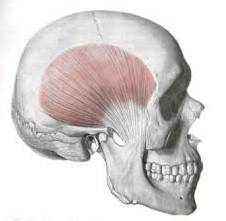
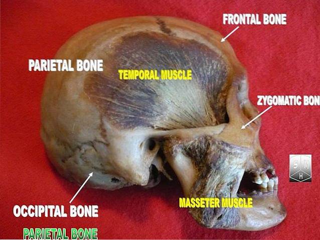
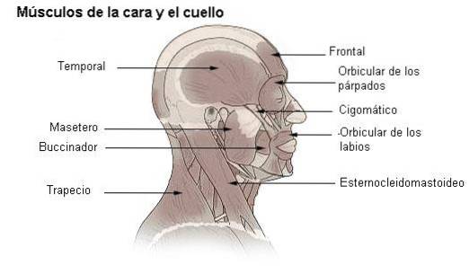
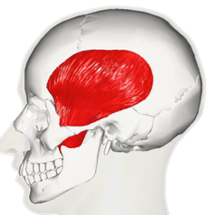
The temporal muscle It is a triangular-shaped paired muscle that is located on each side of the skull, occupying the temporal fossa and a large part of the temporal bone area. This flat muscle is also called the temporalis or crotafies muscle, and is part of the muscle group known as the chewing muscles, due to its powerful action as an elevator of the jaw..
There are pathologies associated with the temporal muscle that are not frequently studied and diagnosed, confused with tension-type headaches and with inflammations in the temporo-mandibular joint.
Article index
The muscle fibers originate superiorly in the temporal bone, in the inferior temporal line above the temporal fossa. Some fibers are also inserted in the entire extension of the temporal fossa located below the aforementioned line.
The fibers are also inserted in the deep face of the temporal aponeurosis, which is the covering aponeurosis, and in the medial region of the zygomatic arch on its internal face by means of an accessory bundle called the jugal bundle..
These multiple insertions make all its fibers, taking as support points different bone structures, act in the elevation of the jaw. Hence, it is the most representative muscle of this movement and allows its combination with protrusion or retraction movements of the jaw..
From here it goes downwards and forwards occupying a large part of the temporal bone area, approximately 70% of it..
Due to its superficiality, it can be palpated without any difficulty when making opening and closing movements of the oral cavity..
Its fibers converge in a strong and resistant tendon that crosses the space between the zygomatic arch and the lateral aspect of the neurocranium, finally inserting itself into the coronoid process of the mandibular bone..
Some fibers also insert into the anterior ramus of the mandible, behind the last molar on each side.
Its main function is to raise the jaw and protrude it forward, thanks to the almost completely vertical fibers of the anterior portion of the muscle.
Similarly, the fibers of the posterior portion, being almost completely horizontal, allow the mandible to move backwards in a protruding movement and to the sides.
In this way, together with the rest of the chewing muscles, they allow the destruction of the food bolus for its subsequent passage to the esophagus.
When talking about the irrigation of the temporal muscle, it is of interest both the irrigation of the muscle itself, and of the fascia that covers it..
The anterior deep temporal artery and the middle deep temporal artery are branches of the maxillary artery, which in turn is one of the terminal branches of the external carotid artery..
Both anterior and middle deep temporal arteries emit branches that are distributed towards the temporal muscle and anastomose with the middle temporal artery..
The middle temporal artery is in turn a branch of the superficial temporal artery, which is another of the terminal branches of the external carotid artery and is responsible for supplying the temporal fascia.
One of the collateral branches of the superficial temporal artery, the posterior deep temporal artery, passes through the temporal fascia and is responsible for supplying the deep aspect of the temporal muscle..
The innervation of the temporal muscle is given by branches of the mandibular nerve, which is the largest and lowest of the three branches of the trigeminal nerve..
The trigeminal nerve is also called the fifth cranial nerve or trigeminal nerve. It is a mixed nerve, that is, it is responsible for both motor and sensory innervation of the structures it innervates, as is the case of the temporal muscle..
The particular case of this muscle is that it receives the innervation of 3 different nerves, one for each anterior, middle and posterior fascicle..
The mandibular branch of the trigeminal nerve provides a temporomandibular trunk from which the anterior deep temporal nerve bifurcates, which crosses the zygomatic foramen just like the temporal muscle and innervates the fascicle or anterior 1/3 of the muscle..
A second trunk of the mandibular branch of the trigeminal nerve gives rise to the posterior deep temporal nerve, which also crosses the zygomatic foramen and reaches the temporal muscle to innervate its posterior fascicle..
Similarly, a collateral branch emerges from the mandibular branch, which is called the medial deep temporal nerve. Like the previous one, it makes its way to the temporal muscle to innervate its middle fascicle.
Temporal muscle syndrome is the most common disease of the temporal muscle, which causes headaches similar to those caused by hypertensive conditions (tension headaches)..
The pain usually appears spontaneously or on palpation over the zygomatic arch and tends to radiate towards the eye or ear.
Usually occurs unilaterally, although it can occur on both sides.
It can be justified by a certain stiffness of the muscle as it is trapped in its passage through the zygomaticus and brings with it loss of stability and vertigo.
Treatment consists mainly of avoiding protrusion movements of the jaw when speaking, chewing, among others. In some cases it is necessary to use an inverted balancer to avoid involuntary movements of this type..


Yet No Comments