
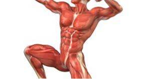
The antigravity muscles or antigravitational are a set of muscle groups whose primary function is to support the force of gravity to maintain a certain posture in the healthy individual. The set of muscle fascicles exerts counter-regulatory functions in favor of a postural axis.
This set acts in a synergistic and harmonious way to overcome the gravitational force and provide stability and balance. The importance of antigravity muscles in their anatomy, physiology and distribution, lies in the fact that their alteration could cause serious repercussions in the lives of individuals due to their involvement in passive and active movements of the body..
Article index
Antigravity muscles differ depending on the type of movement they perform. The different types that exist are described below:
- Antigravity muscles with movements in a downward direction (in favor of gravity).
- Upward motion antigravity muscles (against gravity).
- Horizontal motion antigravity muscles (perpendicular to the force of gravity).
On the other hand, there is also a classification of antigravity muscles that divides them according to the type of action they perform:
- Static muscles, which to be used continuously. They are usually in a state of contraction and are characterized by being the most apt to resist stretching..
- Dynamic muscles, which generate movements from their contraction in the joints. They are more apt to carry out the movements.
Among the various functions that are attributed to the antigravity muscles, the following can be detailed:
- Postural function: it is the most important function of this set of muscles. They are in charge of maintaining a certain posture in the individual who opposes the force of gravity.
- Proprioceptive function: as structures that have proprioceptors, they are capable of sending information from body segments to the cerebral cortex.
- Tonicity function: due to their continuous traction, they are responsible for giving the body the appearance of tonicity.
Muscle that divides the thorax from the abdominal cavity, acting as an anatomical stool. Provides stability and balance to the body, and when it contracts it increases the emptying of the blood found in the liver.
It has various origins because it is composed of numerous fibers that have an anchor or support point in all the anatomical structures that form the lower costal orifice.
It has a frenetic center in the shape of a clover where all its muscle fibers converge.
Muscle located below the oblique of the abdomen. Among its functions are the increase in intra-abdominal pressure and the constriction of the abdomen, which leads to synergy in the processes of expiration, urination, defecation and all those that require an increase in intra-abdominal pressure..
It originates from the medial aspect of the fifth or sixth ribs and the costiform processes of the L1 - L5 lumbar vertebrae.
It inserts on the midline of the body, specifically on the pectineal line, pubic crest and linea alba, thus forming an anatomical structure known as the arch of Douglas..
Muscle located in the posterior part of the humerus, its main function is the extension of the forearm and arm. It is made up of 3 portions: one long, one medial and one lateral.
Its long portion originates from the scapular infraglenoid tubercle, the lateral portion originates above the humeral torsion canal, and the medial portion originates immediately inferior to the humeral torsion canal..
They insert into the olecranon through a common tendon called the triceps tendon..
Muscle located in the lower limb, at the height of the femur; its main function is the extension of the knee. It is the main antigravity muscle, bearing the greatest amount of weight on itself. It is made up of 4 portions: one lateral, one medial, one intermediate and one anterior.
The vastus medialis originates from the intertrochanteric line to the linea aspera of the femur, inserting into the patella.
The vastus lateralis originates in the external and superior part of the femur and inserts in the inferior area of the greater trochanter.
The vastus medialis originates from the upper two thirds of the lateral aspect of the femur, and the rectus anterior originates from the anteroinferior iliac spine and the cotyloid eyebrow.
Together, all portions of the quadriceps femoris unite at the most distal part of the femur, forming a bulky tendon that attaches to the base and sides of the patella.
Muscle whose function is to abduct and rotate the femur.
It has an extensive origin in the lateral border of the iliac crest, the external iliac fossa, the gluteal aponeurosis and the anterior superior iliac spine..
It is inserted on the external aspect of the greater trochanter.
It is a muscle located at the level of the iliac crest with various functions, among which the flexion of the thigh on the pelvis and the recovery of the erect position from the crouched position stand out..
Its origin is in the upper two thirds of the external iliac fossa, in the coccyx, in the sacroiliac ligaments and in the posterior part of the sacrum..
It is inserted in the rough line at the height of its trifurcation.
Muscle whose action is the flexion of the hip.
It has its origin in the transverse process of the lumbar vertebrae and internal iliac fossa.
Lesser trochanter of the femur.
Muscle made up of two bellies. It is located on the thigh and has a triangular shape. Its function is the retroversion of the pelvis, keeping the spine stable. At the level of the femur is adductor and internal rotator.
It originates at the level of the pelvis, in the posterior two thirds of the ischiopubic ramus.
One of its bellies is inserted in the rough line of the femur and the other in the posterior aspect of the medial condyle of the femur.


Yet No Comments