
The leg muscles are the muscles that help us move from one place to another and support our weight when we are standing. They are what allow us to move the foot in relation to the leg and the leg in relation to the thigh when we sit, walk, run and do many other physical activities during the day, every day.
The legs represent the lower limbs of the human body and the bones that make them up belong to the appendicular skeleton, which is the part of the skeleton that includes the bones of both the hands, forearms and arms, as well as the thighs, legs. And feet; and the shoulder girdles (where the shoulders are) and pelvic (where the hips are).
So the legs, with their bones and the muscles that help to move them, belong to the locomotor system. Each leg of our body is made up of about 32 bones, which are associated with a large number of muscles that are between them and the skin that covers them..
To understand a little more the anatomical relationships of the muscles in the lower limbs, it is convenient to quickly mention the bones that compose them. Thus, from top to bottom (from the pelvic girdle to the foot) the bones of the legs are:
Leg muscles, as part of the musculoskeletal system, are striated skeletal muscles, which means that they are made up of long cells that contain contractile proteins arranged in such a way that, when viewed under the microscope, they have a striated appearance..
They are mostly long muscles, capable of stretching a lot. The contraction and relaxation of these is what makes possible the movement of the bones that are part of the legs, which helps us, among other things, to move.
The legs also have short muscles that provide stability to the joints (the contact / junction sites between two or more bones), the ability to rotate and perform "fine" movements, etc..
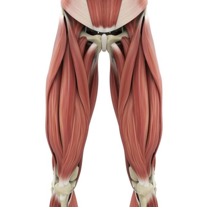
In the thigh are the strongest muscles in our entire body, since they are the ones that have the responsibility of extending the knee so that it has a straight position, thus allowing the daily movement of the legs, either to walk, climb or descend stairs lift weight, etc.
These muscles, then, are the ones that support most of our body weight, which makes them very important to us..
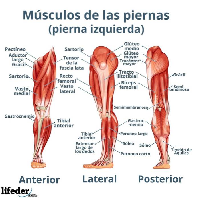
The muscles of the anterior region of the thigh participate in flexion and lateral rotation of the hip, while the muscles of the posterior part extend, abduct and rotate the hip moderately.
The hips are associated with different types of muscles as well, which are divided into two regions, an anterior (in the same plane as our face) and a posterior (in the plane corresponding to our back or our back).
In the posterior region of the hips are the muscles known as glutes; These are arranged in three layers and are known as:
The anterior region of the thigh is provided with the most colorful and largest muscles in our body: the sartorius muscle and the quadriceps, which is actually made up of 4 individual muscles: three of them superficial and one hidden..
In the anterior region there are also the muscles of the adductor group, which facilitate the movements of the leg contrary to those of abduction, that is, they allow us to close the legs. These muscles are as follows, and are located between the femur and the pubic bone of the hip:
There are three main muscles in the posterior thigh region, which are commonly known as hamstring muscles. These and other important muscles in this region are:
Although we know the leg as everything that is included between our hips and our feet, the region properly known as the leg is the one that encompasses the space between the knee and the foot, that is, where we have the calf.
These are some of the most important muscles in the lower part of our legs, specifically those in the anterior region:
The superficial muscles of the posterior region of the leg are:
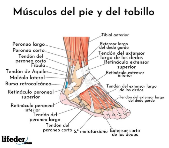
The muscles of the foot are classified as extrinsic (if they are derived from the muscles of the lower leg) or intrinsic (if they are located specifically in the region of the foot).
The extrinsic muscles of the foot function in general movements of the foot such as flexion, rotation, inversion, etc. The intrinsic muscles are responsible for the "fine" actions of the feet, such as the movement of individual toes, for example..
Dorsal region:
Plantar region:
This region is made up of 10 muscles separated into 4 "layers". The muscles in this part of our feet are very important, as they help us control the movement of the toes and stabilize the arch region of the foot..


Yet No Comments