
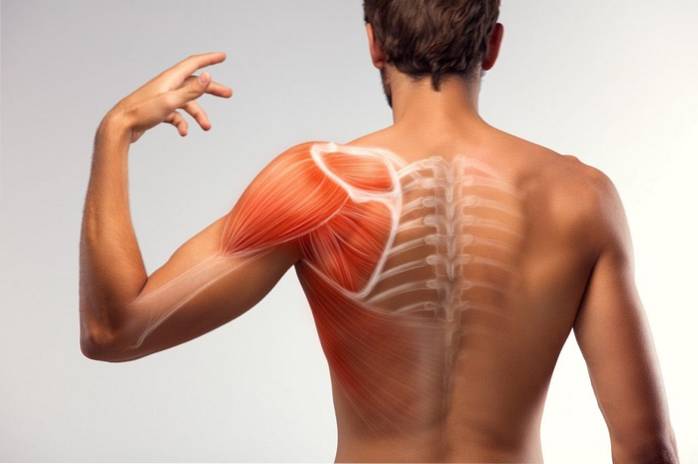
The shoulder muscles are those that are associated with this joint that connects the bones of our arms with the rest of the trunk, that is, part of our appendicular skeleton with our axial skeleton.
The shoulders are one of the most complex joints in our body and, like many others, they are made up of bones, ligaments, tendons and muscles..
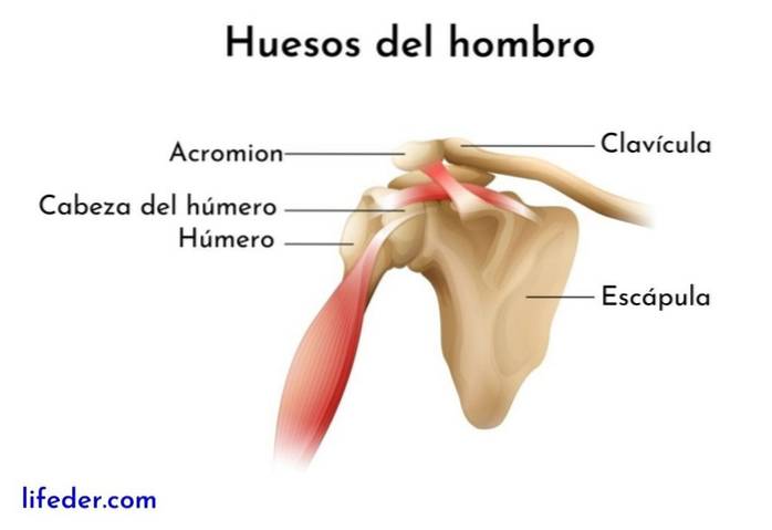
Each shoulder consists of the union between the arm bone (humerus), the scapula (shoulder blade) and the clavicle, the latter belonging to the so-called “shoulder girdle”, responsible for joining the arm with the thorax.
The muscles associated with this important joint allow us to have a wide range of movements and, fundamentally, protect the main junction between the humerus and the scapula, also known as glenohumeral.
Our shoulders have more than 10 muscles that work together to stabilize the joint and control the different movements of rotation, abduction, adduction, flexion and extension that it can perform..
These muscles are generally categorized, according to their position, into two groups: one anterior and one posterior. Let's see what they are, where they are and what functions they fulfill:
These are known as the axioappendicular anterior muscles, as they are located in the front or anterior part of the shoulder. These can also be referred to as thoraco-appendicular muscles, given their function of connection between the thorax and our upper limbs (the arms)..
To this group belong 4 muscles: the pectoralis major, the pectoralis minor, the subclavian and the serratus anterior..
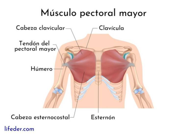
It is a large muscle that covers the anterior region of the rib cage. It is shaped like a fan and extends from the collarbone to the center of the chest.
It has three heads: a clavicular, which arises in the middle part of the clavicle; a sternocostal, which arises from the sternum and the first 6 ribs, and an abdominal one, which arises from the rectum sheath, which corresponds to the lining of the anterior abdominal muscles.
Participates in the adduction and mid-rotation movements of the arm, as well as in the inhalation during breathing. These are the movements we make when we lift our arm and "open" it or project it towards the front of our chest..
It is a smaller, thinner and flattened muscle that is located below the pectoralis major muscle..
Your job is to connect the shoulder joint, specifically the scapula, with the third, fourth, and fifth rib; allows the movement of "projection" of the shoulder blade and also helps us lower our shoulders.
This muscle arises from the cartilage of the first rib and attaches to the middle part of the anteroinferior surface of the clavicle..
It is a muscle that is divided into three parts: upper, middle and lower.
The upper part arises from the first two ribs and attaches to the upper angle of the anterior aspect of the scapula..
The middle part arises between the third and sixth rib and inserts on the middle edge of the scapula, while the lower part arises between the seventh and ninth rib and inserts on the edge and lower angle of the scapula.
This muscle participates in the anterolateral movement of the scapula, as well as in its rotation and its union with the thorax..
The muscles in this group are called “posterior axio-appendicular muscles” or scapulohumeral muscles, since they are the ones that allow the connection and movement between the long bone of the arm (the humerus) and the scapula or shoulder blade..
In this group, two subgroups are distinguished: the intrinsic muscles and the extrinsic muscles..
The intrinsic muscles of the posterior group are 6: the deltoid, the teres major and 4 important muscles of the “rotator cuff”, which are the supraspinatus, the infraspinatus, the teres minor and the subscapularis..
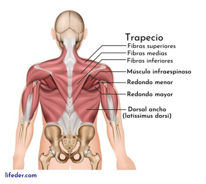
There are 5 extrinsic muscles of the posterior group: the trapezius, the latissimo of the back, the levator scapulae, the rhomboid major and rhomboid minor..
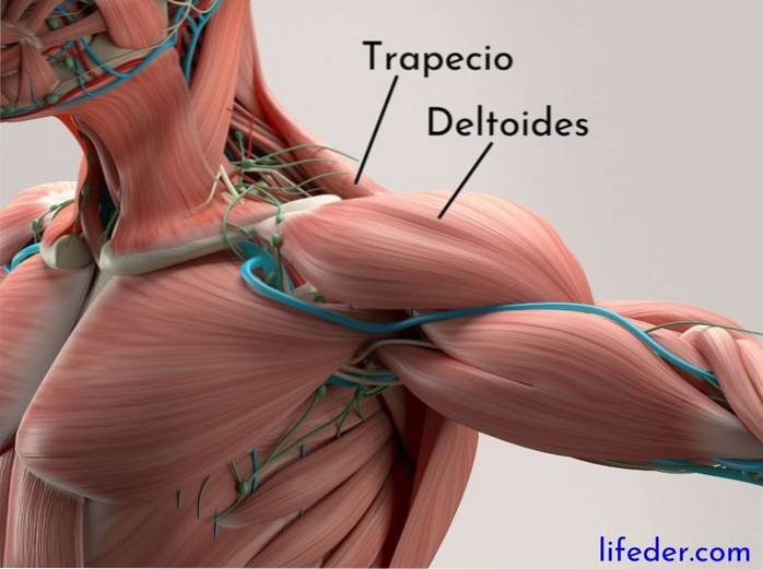

These are very important muscles for the shoulder joint. Basically they are those that surround the glenohumeral joint, forming a kind of bracelet around it..
Two muscle "layers" are generally distinguished, one superficial and one deep. The superficial is formed by the trapezius and the latissimo of the back, while the deep is made up of the levator scapulae muscle and the rhomboid major and minor muscles..
It is a very large muscle that has three parts: ascending, transverse and descending that, together, form the region of the neck..
Each one of the portions of this great muscle exerts a certain function: the descending one participates in the lateral flexion of the head and the neck, as well as in the rotation of the head; the transverse one allows lateral movement of the scapula and the ascending one does it inferomedially.
It is another large muscle found in the middle of your back. It has a vertebral part that arises in the last thoracic vertebrae (lumbar region), another iliac part that arises in the posterior crest of the ileum, a costal part that arises in the 9-12 ribs and a scapular part that arises in the lower angle of scapula.
All parts converge at the same point of insertion: the inter-trabecular groove of the humerus. This muscle participates in the movements of adduction, internal rotation and arm extension, but they also play an important role during breathing..
This muscle arises from cervical vertebrae 1-4 and attaches to the middle edge of the scapula, expanding from the upper angle of the scapula. Works on lateral neck flexion, as well as inferior rotation of the glenoid cavity, associated with the glenohumeral joint.
There are two rhomboid muscles: the rhomboid major and the rhomboid minor. The minor arises in the ligament of the neck and the processes of the vertebrae C7-T1, inserting in the root of the spine of the scapula.
The rhomboid major arises in the processes of the T2-T5 vertebrae and inserts on the medial border of the scapula. Both muscles share functions, since they participate in the supero-medial movement of the scapula and in the rotation of the glenoid cavity.


Yet No Comments