The trunk muscles are all those muscular structures that cover the thorax and abdomen, providing protection and movement to the thoracoabdominal structures. Most of these muscles are flat and wide, covering a large amount of space and presenting bony attachments in more than two points of the skeleton..
They also have the particularity of overlapping with each other, forming a kind of framework, especially in the anterior abdominal wall, where there is less bone support..
Apart from the flat and wide muscles that literally make up the thoracoabdominal walls, there are also a series of long and narrow muscles, most of them attached to the spine or located between the ribs..
These muscles are powerful and have multiple attachments to the vertebrae, which are responsible for keeping the back upright. In addition, they allow flexion-extension and rotation movements of the spine.
Article index
The muscles of the trunk can be classified according to their insertions in:
- Muscles that attach exclusively to bony structures in the thoracoabdominal region.
- Muscles where part of the attachments are in the thoracoabdominal region and part in other anatomical regions (upper limb, lower limb or neck).
On the other hand, these muscles can be classified according to the size and arrangement of their fibers into wide and flat muscles, and long and narrow muscles..
Most of the muscles of the thoracoabdominal wall that are part of the trunk wall can be approached more or less easily from the surface, with one exception: the diaphragm..
The diaphragm is a large, flat, wide muscle located within the thoracoabdominal cavity. In fact, it constitutes the physical boundary between the thorax and the abdomen. The function of the diaphragm is to allow movement of the chest for breathing, in addition to physically separating the abdominal and thoracic compartments.
These are the muscles of the thoracoabdominal wall proper. They are arranged in superficial and deep planes both in the posterior wall (back) and in the anterolateral wall of the thoracoabdominal region..
Among all the muscles that are inserted exclusively in thoracoabdominal bone structures, the diaphragm deserves special mention, since it is the only one that is located entirely within the thoracoabdominal cavity. In addition, it is the muscle responsible for respiration.
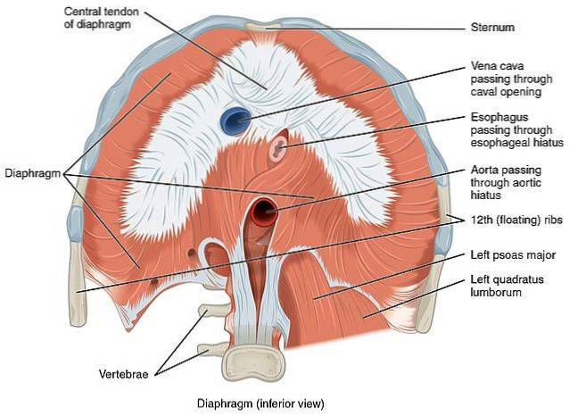
It is a large, broad, flat muscle that, like a dome, forms the floor of the thorax and the roof of the abdomen. Forms attachments in the dorso-lumbar spine, the last costal arch and the sternum.
It is a powerful muscle, responsible for breathing. It has the peculiarity of being an involuntary muscle that can be controlled.
Unlike the heart, which beats without the individual's will, the diaphragm exerts its function (respiratory movements) automatically; but with the difference that there is a certain voluntary control over it. This makes it a unique muscle in the entire body..
In addition to its respiratory function, the diaphragm serves as an anatomical boundary and barrier between the structures of the thorax and those of the abdomen, helps to maintain the pressure differential between both compartments of the trunk and also has openings that allow the passage of the corresponding structures from the trunk towards the abdomen.
It is therefore the most complex and important muscle in the thoracoabdominal region, since it is responsible for respiration, one of the vital functions of the body..
This group includes all the erector muscles of the spine, which are located throughout the entire back. Each of them is thin, of variable length (there are short and also very long); and usually form multiple insertions in vertebral processes.
The erector spinal muscles overlap each other just like the links of a chain, and allow a great range of motion both flexion-extension and rotational to the spine.
These muscle groups include the following muscles:
- Interspinous muscles.
- Trasverse-spinous muscles.
- Intertransverse Muscles.
All of them run cephalocaudal and are located in the midline of the back covered by an intermediate muscular plane.
At the level of the thorax, there are no deep muscles outside the midline, this space being occupied by the ribs and the intercostal muscles..
In the abdomen, the oblique muscles of the abdomen are occupying the deep plane and outside the midline. These large, wide and long muscles “wrap” the abdominal wall taking insertions from behind in the spine, above in the last costal arches, and below in the pelvis..
The abdominal muscles are part of the deep plane of the posterior abdominal wall, as they are covered by other muscle planes. However, in the anterolateral wall of the abdomen they become superficial, since they are not covered by other muscular structures..
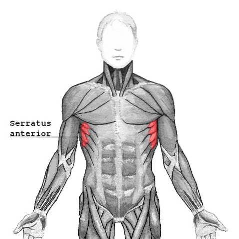
The medial plane is made up of muscles that take attachments to the scapula. From there they extend, either to other bony structures of the thorax, or to the upper limb.
The muscles that take insertion both in the scapulae and in the vertebral bodies or ribs are the following:
- Rhomboids major.
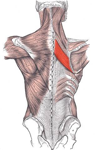
- Minor rhomboids.
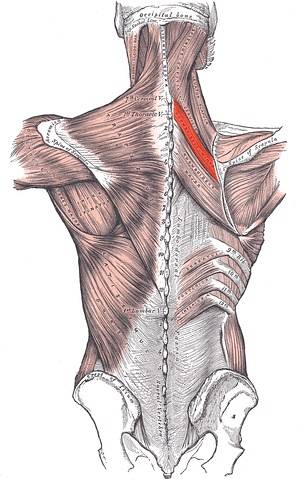
- Serratus anterior.
The rhomboid muscles take insertion at the medial border of the scapula, and from there they go towards the spinous processes of the dorsal vertebral bodies..
For their part, the serratus take insertion on the same edge of the scapula but in a deeper plane, passing under it. Later they move forward on the anterolateral chest wall to insert into the costal arches.
The muscles that take attachments to both the abdominal wall and the arm are described later..
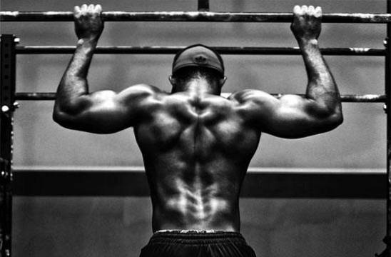
This group is made up of two large muscles: the trapezius and the latissimus dorsi..
Both muscles occupy the superficial part of the back, overlap each other and cover the entire posterior region of the trunk, from the sacrum to the head. 95% of their extension is on the trunk, although they have distal attachments in the neck (trapezius) and upper limbs (latissimus dorsi).
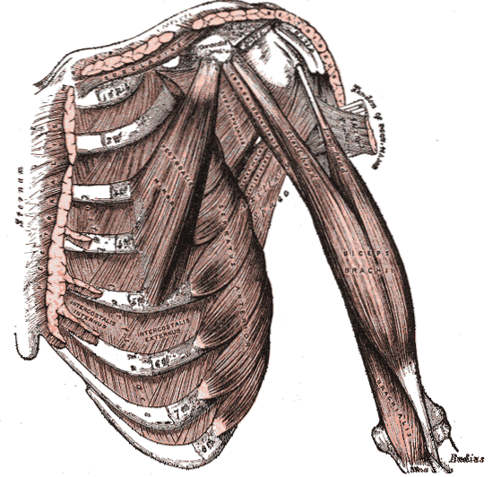
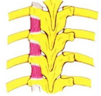
They are small, narrow and short muscles that are located between the ribs, taking insertion in both the upper and lower rib to each of them.
In each intercostal space there are three intercostal muscles, namely:
- External intercostal.
- Internal intercostal.
- Middle intercostal.
The external intercostal muscle is the most superficial of the three and is located in the entire extension of the intercostal space, seat of the rib tubercles up to the costochondral junction..
For its part, the internal intercostal is the deepest, and it locates approximately the anterior two thirds of the costal arch (it does not reach the back). Its fibers usually extend from the sternum to the costal angle.
In the area where the intercostal vessels cross the internal intercostal, it presents a split into two muscle bellies, one internal (internal intercostal) and the other more superficial. The latter is known by some authors as the middle intercostal.
The intercostal muscles are found in the thickness of the thoracic wall, covered in the posterior region by the muscles of the median and superficial planes of the back and in the anterior region by the pectoral muscles.
Only in the lateral region are they easily accessible, being covered exclusively by subcutaneous cellular tissue and skin. Due to this particularity, this is the site of choice for the placement of chest drainage tubes..
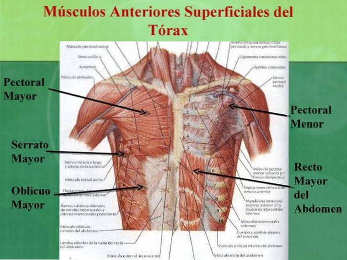
The muscles of the anterior region are the subclavian (which joins the clavicle with the first costal arch) and the pectoralis major and minor..
The pectoralis minor can be considered a proper muscle of the trunk, since it goes from the coracoid process of the scapula to the first three ribs. It is located immediately in front of these, forming the deepest plane of the pectoral region.
Immediately above this and covering it in its entirety is the pectoralis major. As with the latissimus dorsi and trapezius, 90% of the muscle mass of the pectoralis major is found covering the anterior thoracic wall, although it also takes insertion in the humerus.
They are powerful and robust muscles that not only provide mobility to the arm but also protection to the rib cage and support to the overlying structures. This is especially true in women, where the mammary gland is closely related to the pectoralis major through the clavideltopectoral fascia..
The muscles of the anterolateral region of the abdomen are, as already indicated above, the abdominal muscles.
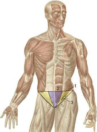
The lateral abdominal wall is made up of three broad muscles, overlapping and overlapping each other:
- Major oblique.
- Minor oblique.
- Transverse abdomen.
The greater oblique is the most superficial of the three and covers all of them. Its fibers run from top to bottom and from outside to inside.
Immediately below this is the minor oblique muscle. Its fibers go in the opposite direction, from bottom to top and from back to front. Finally, in the deepest plane is the transverse abdominal muscle, whose fibers run perpendicular to the major axis of the body..
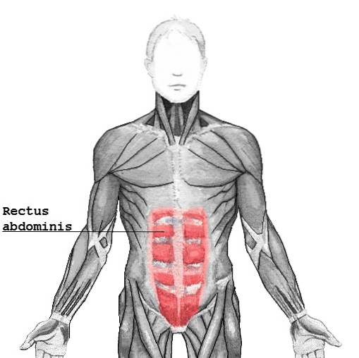
The abdominal muscles take multiple attachments into the spine from behind, the last costal arches (10, 11, and 12) above, and the pelvis below. Towards the anterior wall, the aponeurosis of all of them condenses to merge with the sheath of the rectus abdominis muscle, the only one located in the midline of the anterior wall.
The rectus abdominis muscle is wide, flat, and thick. It occupies the midline and takes proximal attachments in the 10th costal arch and xiphoid appendix, while its distal attachments lie on the symphysis pubis.
In the midline the anterior rectus abdominis left and right merge into an aponeurotic thickening known as the linea alba..
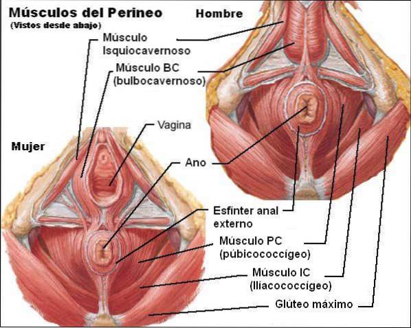
They are short, strong little muscles that make up the floor of the pelvis. They are classically described as a separate region (perineum), but functionally they constitute the floor of the entire abdominal cavity. Therefore, they should be mentioned when counting the trunk muscles..
This region includes the following muscle groups: superficial plane, median plane, and deep plane..
- External sphincter of the anus.
- Superficial transverse perineum.
- Ischiocavernosus.
- Bulbocavernosus.
- Vulvar constrictor muscle.
- Deep transverse perineum.
- Urethrovaginal sphincter.
- Urethra compressor.
- Levator ani.
- Ischiococcygeus.
- Pubococcygeus.
Most of these muscles are located in the posterior region of the trunk, forming the intermediate muscular plane in the dorsal region of the thorax..
They are powerful muscles, which connect the upper extremities with the trunk, for which they take insertions both in the thoracoabdominal bone structures and in the axial skeleton.
These muscles include the following:
- Infraspinous.
- Supraspinatus.
- Major round.
- Minor round.
- Subscapular.


Yet No Comments