
The pronator muscles are two muscles responsible for tilting the radius bone through the ulna bone or in a prone (face down) position.
There are two types of pronator muscles: the pronator teres muscle and the pronator quadratic muscle..
The word pronator comes from the Latin Pronusm which means "leaning forward or lying face down." Pronation is a movement of the hand, wrist, and forearm, which is unique to the human body..
While rotating at the top during supination, the radius bone circles half a circle at its distal end over the ulna bone as its center point. Basically the radius crosses over the ulna bone, when the hand and wrist move from the palm down.
The pronator muscle originates from the medial epicondyle of the humerus and inserts on the axis of the radius. By traveling with the bone radius half its distance before inserting it, the pronator muscle can take advantage of the leverage.
This means that half of the radius of the bone is used as a lever to roll over the ulna bone, giving one the ability to pronation with the power of the elbow. A capacity that comes from the pronator quadratic muscle.
The pronator muscles are innervated by the median nerve. When the pronator muscles spasm, they make pronation weak and supination restricted.
The pronator teres muscle, also known as the pronator teres, is a muscle of the human body that is mainly found in the forearm, and that together with the pronator square, serves for the pronation of the forearm.
It is the stronger of the two pronator muscles, however, it is only activated during fast or resisted forearm pronation. The pronator teres has two heads: humeral and ulnar.
The humerus head, the largest and most superficial, arises from the medial supracondylar ridge immediately superior to the medial epicondyle of the humerus and from the common flexor tendon (arising from the medial epicondyle).
The ulnar head is a thin bundle, arising from the medial side of the coronoid process of the ulna, and joins the anterior at an acute angle.
The pronator teres has a tendency to be hyperactive and short due to overuse. This abuse can be caused by repetitive activities that involve a pronated forearm position or active forearm pronation movement, including throwing, some strokes such as when playing tennis, swinging a golf club, and turning a screwdriver or wrench..
Also, exercises that involve holding the forearm in a pronated position and isometrically contracting the pronator can contribute to its overuse..
As the pronator becomes shorter and shorter, the tension across the muscle increases and the quality of the tissue deteriorates, often leading to injury..
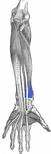
The pronator square is a rectangular muscle in the front of the forearm that connects between the radius and the ulna..
It is the main promoter of forearm pronation, receiving help from the pronator teres during rapid pronation. It is also known for its role in preventing separation of the ulna and radius when force is transferred to the forearm through the heel of the palm of the hand..
Classified as part of the deep anterior compartment of the forearm, the pronator quadratus is the deepest of the muscles in the front of the forearm, and is deeply embedded in the mass of the flexor tendons of the wrist..
Its parallel muscle fibers extend laterally from their origin in the distal anterior ulna. The fibers cross over the interosseous membrane of the forearm before inserting into the distal anterior ulna, forming a flat square muscle shape..
The pronator square can become hyperactive and short due to overuse of repetitive activities that involve the forearm pronation movement, as well as activities that involve excessive isometric contraction of the pronator muscles..
Some syndromes that can affect the pronator muscles are:
Carpal tunnel syndrome is a common condition that causes a tingling sensation, numbness, and sometimes pain in the hand and fingers. These sensations develop gradually and usually begin to worsen at night. They tend to affect the thumb, index finger, and middle finger.
Pronator teres syndrome (also called pronator syndrome) is a compression neuropathy of the median nerve in the elbow..
It is not as common as compression in the wrist, which is carpal tunnel syndrome. It occurs more often in women over 40 years of age.
Medial nerve compression at the elbow can cause pain and / or numbness in the distribution of the distal median nerve, and weakness can develop in the flexor of the long finger of the thumb and the deep flexor of the index finger and pronator quadratic.
Symptoms include tenderness over the pronator teres and pain with pronation of the resisted forearm. Weakness could be present with thumb abduction, as well as deterioration of the pincer muscles. Sensation changes can also be experienced in the first three fingers and the palm.
Anterior interosseous nerve syndrome is a rare syndrome comprising less than 1% of all upper extremity nerve palsies. It is so called because it arises from compression or inflammation of the anterior interosseous nerve of the forearm.
This syndrome innervates three muscles in the forearm: the pronator quadratus, the long flexor of the thumb, and the radial half of the deep flexor of the finger..
Most people with this syndrome feel localized pain in the forearm. The pain is sometimes described in the ulnar fossa and mainly causes pain in the elbow. What is characteristic is the deterioration in the movement of the thumb and index finger..


Yet No Comments