
The spinal or spinal nerves They are those whose origin is in the spinal cord and that reach different areas through the vertebral muscles. They belong to the somatic nervous system, and include a total of 31 pairs of nerves that innervate the entire body except for the head and some parts of the neck..
Of the 31 pairs that make up the set of spinal nerves, there are eight cervical, twelve dorsal, five lumbar, five sacral, and a coccygeal pair. Furthermore, they all have a mixed function; that is, they are both sensitive and motor, carrying information both from the spinal cord and towards it..

The spinal nerves are numbered from top to bottom, naming them according to the body region in which they are located. The two roots of each of them have their origin in the spinal cord, having a sensitive posterior and an anterior motor. Both join to form the trunk of the spinal nerve, which passes through an intervertebral foramen.
In this article you will discover all the existing information about the 31 pairs of spinal nerves. In addition, we will also study everything we know today about its functions, and we will see more about its anatomy and location..
Article index
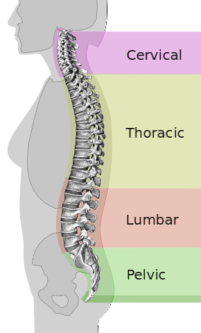
The spinal nerves are divided into five groups. Each of them is related to an area of the spine, and their names have their origin in the vertebrae from which they arise. Next we will see in detail each one of them.
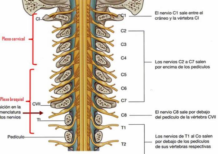
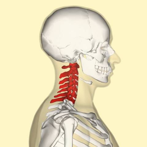
The cervical nerves are those spinal nerves whose origin is in the cervical segment of the spinal column. Although there are only seven cervical vertebrae (C1 - C7), there are eight nerves of this type (C1 - C8).
All of them except C8 arise above its corresponding vertebra, the latter emerging below C7.
This makes them different from the rest, since the others emerge below the vertebrae that give them their names. The posterior distribution includes the suboccipital nerve (C1), the greater occipital nerve (C2) and the third occipital nerve (C3).
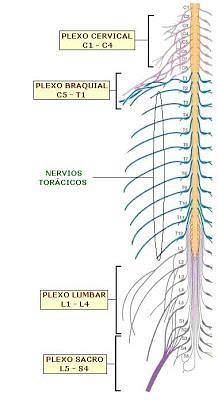
On the other hand, the anterior distribution includes the cervical plexus (C1 - C4) and the brachial plexus (C5 - T1). The cervical nerves, on the other hand, innervate muscles such as the sternohyoid, the sternothyroid, and the omohyoid..
The thoracic nerves are the twelve spinal nerves whose origin is found in the thoracic vertebrae. Each of them comes out below its corresponding vertebra: thus, the T1 has its origin under the one of the same name. Its branches go directly to the paravertebral ganglia, which are part of the autonomic nervous system..
The thoracic nerves are involved in the functions of organs and glands in the head, neck, chest, and abdomen. On the other hand, there are several divisions that are important to consider when studying them..
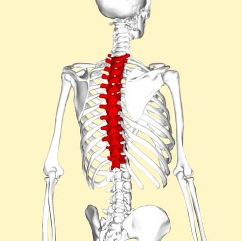
In the anterior divisions, the intercostal nerves come from nerves T1 through T11, and pass between the ribs. In T2 and T3, other branches form the intercostobrachial nerve. The subcostal nerve arises from T12, and passes under the twelfth rib.
Regarding the posterior divisions, the medial branches of the posterior branches of the six superior thoracic nerves pass between the semispinal dorsum and the multifidus. Afterwards, they reach the rhomboid and the trapezius, and reach the skin on the sides of the spinous process. This sensitive branch is known as the medial cutaneous branch..
The medial branches of the six inferior thoracic nerves are distributed mainly towards the multifido and the longissimus dorsi, although occasionally some of their filaments reach the skin. This sensitive branch is known as the posterior cutaneous branch..
The lumbar nerves are those that emerge from the lumbar vertebrae. They are separated into posterior and anterior divisions.
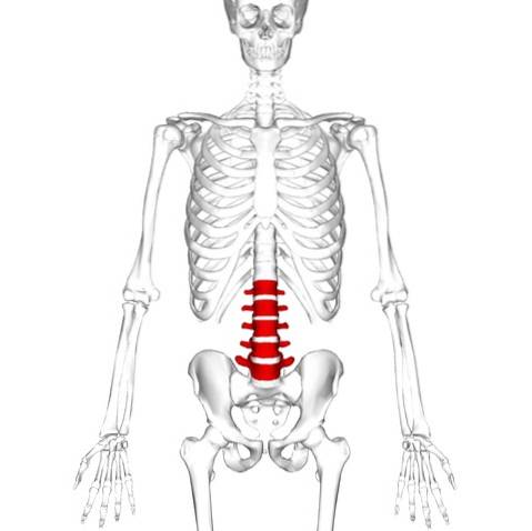
The medial branches of the posterior divisions of the lumbar nerves pass close to the articular processes of the vertebrae and terminate in the multifidus muscle. The laterals work in conjunction with the erector spinae muscles.
The three superior lumbar nerves send cutaneous nerves to the latissimus dorsi at the lateral border of the erector spinae muscles. Later, they descend through the back of the iliac crest, until they reach the skin of the buttocks. Some of its ramifications extend to the level of the great trochanter.
The anterior divisions of the lumbar nerves and their branches increase in size the further down the body they are. They unite, close to their origins, with the gray rami communicantes of the lumbar ganglia and the sympathetic trunk.
These rami are formed by long, thin branches that accompany the lumbar arteries around the sides of the vertebral bodies, below the psoas major. This arrangement is somewhat irregular, in the sense that one ganglion can branch to two lumbar nerves, or one of these nerves can receive branches from two ganglia..
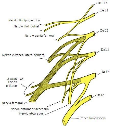
The first four lumbar nerves are connected to the lumbar part of the sympathetic trunk by a white ramus communicans. The nerves pass obliquely outward under the psoas major, or between its fascicles, distributing filaments to both it and the quadratus lumborum.
The first three nerves and much of the second are connected at this location by anastomotic loops, thus forming the lumbar plexus. The smallest part of the fourth joins with the fifth to form the lumbosacral trunk, which aids in the formation of the sacral plexus..
Thus, the L4 nerve is known as the furcal nerve, because it is divided between the two plexuses.
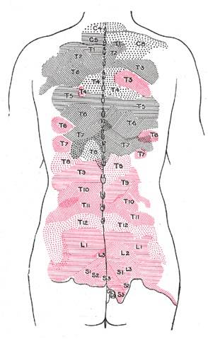
The sacral nerves are five pairs of spinal nerves that exit through the sacrum at the bottom of the spine. Its roots begin within the column at the level of the L1 vertebra, where the cauda equina begins; and later they descend to the height of the sacrum.
There are five pairs of sacral nerves, half of which emerge from the sacrum on the left side and the other half on the right. Each of them emerges in two different divisions: one does so through the anterior sacral foramina, and the other through the posterior sacral foramina.
These nerves are divided into different branches; and those of each of them join with those of the others, as well as with the branches of the lumbar nerves and the coccygeus. These nerve anastomoses form the sacral and lumbosacral plexus. The branches of these plexuses are those that work in areas such as the hips, calves, legs or feet..
The sacral nerves have both afferent and efferent fibers; and therefore are responsible for most of the sensory perception and movements of the lower extremities of the human body.
From nerves S2, S3, and S4 arise the pudendal nerve and parasympathetic fibers, whose electrical potentials work with the descending colon, rectum, bladder, and genital organs. These pathways also have both afferent and efferent fibers; and therefore, carry both sensory information to the CNS and motor commands to these organs.
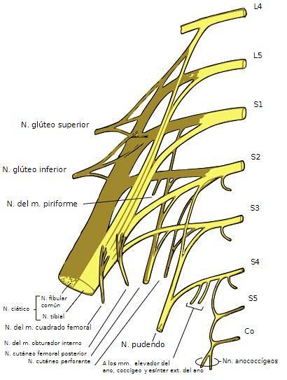
Lastly, the coccygeal nerve is number 31 within the spinals. It arises from the medullary cone, and its anterior root helps to form the coccygeal plexus.
Unlike the previous ones, it is not divided into a medial and a lateral branch. Its ramifications mainly reach the skin on the back of the coccyx.
Spinal nerves travel from the Central Nervous System (CNS) to practically every corner of the human body. With the exception of some areas of the head and neck, which are controlled by the cranial nerves, all the organs, muscles and glands of the body transmit and receive their information through these nerves..
Thus, a single nerve can transmit and collect information from several different organs, from the skin, or from different glands. Through the branches into which they are divided, each of them can perform multiple functions, forming a complex system that connects all parts of the body with the central nervous system..
As we have already seen, spinal nerves are both afferent and efferent. This means that each one of them fulfills a double function; both are essential for the proper functioning of the human body.
On the one hand, the spinal nerves collect information from the organs, glands or muscles with which they are connected and transmit it to the central nervous system through the spinal cord. In this way, the brain can process all this data and elaborate an appropriate response to a certain situation..
On the other hand, the same spinal nerves are responsible for carrying the response produced by the CNS to the effector organs, in such a way that we can react and function correctly in our environment.


Yet No Comments