
The neurulation it is a fundamental phase in embryonic development in which the neural tube develops, a structure that will give rise to the brain and spinal cord (central nervous system).
It occurs in all vertebrate embryos, although in certain species it goes through two different processes: primary and secondary neurulation. The neurulation process begins around the third or fourth week of embryonic development.
The development of our brain is mediated by genetic instructions, intercellular signals, and our interaction with the external world. Initially, this development consists of the establishment of a primal nervous system.
Thus, it begins with the generation of neurons from undifferentiated cells, the formation of main brain regions, and the migration of neurons from their places of creation to their final places. This will lay the foundations for the subsequent creation of axonal pathways and establishment of synapses (connections)..
Article index
To understand the neurulation process, it is necessary to know some fundamental previous steps in embryonic development..
Before the cells that are to become the brain and spinal cord appear, there are primitive cell layers that are essential for the subsequent development of the nervous system. These layers are formed during the so-called "gastrulation", which, as Lewis Wolpert pointed out in 1986:
“It is not birth, not marriage, not death. Gastrulation is really the most important moment of your life ".
During this delicate period, in which a single sheet of cells is divided into the three primitive layers or germ layers:
- Ectoderm or outer layer: gives rise to the epidermis and related structures such as hairs and nails, as well as the nervous system.
- Mesoderm or intermediate layer: from it the muscles, bones, circulatory system, and reproductive and excretory organs will appear.
- Endoderm or inner layer: it will give rise to the digestive system and the respiratory system.
The mesoderm and endoderm invaginate (fold over themselves), defining the midline and the anterior-posterior and dorsal-ventral axes. These axes are important because in each area of the germ layers different events will happen.
Gastrulation also has a key function, which is the formation of the notochord. It begins to emerge at 18 days of gestation, and consists of a defined cylinder of mesoderm cells that expand along the midline of the embryo.
The notochord is formed through cellular movements that occur during gastrulation. At first, a superficial slit is formed called the primitive pit, which is lengthening until it constitutes the “primitive line”. From there the mesoderm invaginates and extends inward to form a cylinder.
The notochord establishes the midline of the embryo, which will result in both halves of the body being symmetrical. This structure also defines the position of the nervous system and is essential for posterior neural differentiation..
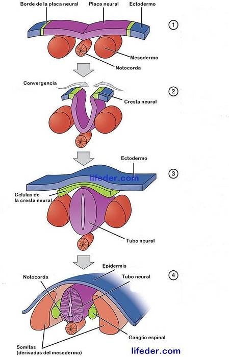
In this way, the neurulation process begins. The notochord begins to send inductive signals to the ectoderm (which is just above it) so that a group of neuroectodermal cells differentiate into nerve precursor cells. The latter are the ones that will be part of the central nervous system..
The part of the ectoderm that covers the notochord is defined as the “neural plate”. As neurulation progresses, the neural plate begins to thicken, accumulating cells. These cells are arranged in two chains on either side of the midline of the neural plate.
The latter begins to fold in the midline (adjacent to the notochord). This gives rise to the neural sulcus, approximately at 20 days of gestation, which is becoming more and more accentuated..
The part of the neural plate that is immediately above the notochord is called the "floor plate." Whereas, the posterior part of the protruding ends of the sulcus is known as the "neural crest".
Little by little, the two protruding cell chains of the neural plate are bending, seeking to touch. This results in a cylinder called the neural tube. The neural tube closes and is completed at approximately 22 days of pregnancy.
The mesoderm that is next to the neural tube becomes thicker, dividing into structures called "somites". These structures are the precursors of the musculature and the skeleton..
During neurulation, different parts of the neural tube will develop different structures in our body. These changes begin at 24 days of gestation. Thus:
- The part of the neural tube adjacent to the somites begins to develop into the rudimentary spinal cord.
- The area of the neural crest will give rise to the sensory ganglia of the peripheral nervous system.
- The anterior ends of the neural plate, called the "anterior neural fold," will expand together in the midline to originate the brain..
- The cavity of the neural tube will become the ventricular system.
Thus, the neural tube will give rise to the brain and the spinal cord. The cells of the neural tube are known as neural precursor cells, which are stem cells from which more precursors will emerge that give rise to neurons and glial cells..
On the other hand, some subsets of neural precursor cells do not divide. They are called neuroblasts, and they will differentiate into neurons.
While the cells of the ventral part of the neural tube (where the floor plate is) go to give rise to the spinal cord and the back of the brain.
At 25 days of gestation, 3 basic vesicles can be seen that start from the neural tube: the forebrain, the midbrain and the rhombencephalon.
While, at 32 days, they are divided into 5 structures:
- The telencephalon: which gives rise to the cerebral cortex, the striatum, the limbic system and part of the hypothalamus.
- The diencephalon: what will develop the epithalamus, thalamus and hypothalamus.
- The midbrain: which will give rise to the brain tectum, tegmentum and peduncles.
- The metancephalon: what will differentiate into cerebellum and brain bridge.
- The myelencephalon: what will become the brainstem (medulla oblongata).
Primary and secondary neurulation are two fundamental phases in the neurulation process. In general, they define two types of neural tube formation.
The anterior part of it will be formed through primary neurulation and the posterior part, by secondary neurulation. Both occur at the same time, but in different places.
Each organism uses different degrees of primary and secondary neurulation; except fish, which use only the secondary.
Much of the neural tube develops during the third week of gestation from primary neurulation. Its formation includes up to somite 31, which gives rise to the second sacral vertebra of the spine.
It begins when the cells of the neural plate begin to proliferate and to be located in two chains separated by an invagination in the midline.
Finally, the chains bend and join, constituting part of the neural tube. This part gives rise to almost the entire nervous system (brain, cervical, thoracic and lumbar spinal cord)..
The rest of the neural tube is formed by secondary neurulation. It arises from the condensation, differentiation and degeneration of the mesenchymal cells that are in that area. (Chávez-Corral, López-Serna, Levario-Carrillo, & Sanín, 2013).
This occurs in the absence of the ectodermal germ layer or neural plate. It begins with the formation of a medullary cord due to the condensation of mesenchymal cells, which is hollowed out to give rise to the neural tube.
This tube, also called the medullary tube, arises from an undifferentiated mass of cells called the causal eminence. Through morphogenetic mechanisms, they will organize themselves forming a cavity to give rise to the spinal cord of the sacral and coccygeal region..
After secondary neurulation is complete, it joins the most caudal part of the primary neurulation.
It is possible that alterations may arise during neurulation due to genetic mutations or other reasons. Around 5 or 6 weeks of gestation, most of the brain and face begin to form. The hemispheres differentiate and the optic vesicles, olfactory bulbs and cerebellum grow.
If this important moment in neurodevelopment is altered, severe neurological and neuropsychological disorders usually appear. These are usually accompanied by seizures..
Alterations in this process lead to serious conditions. Especially if there are defects in the closure of the neural tube, which are not usually compatible with life. These occur between 1 in every 500 live births. The most common disorders that appear due to a bad closure of the neural tube are:
It occurs due to poor closure in the anterior part of the neural tube during neurulation. It is characterized by the absence of some parts of the skull, brain and facial malformations, as well as heart problems.
It arises from a neural tube defect that results in incomplete development of the brain, spinal cord, or meninges (protective layers that surround the central nervous system). There are several types of spina bifida: it can be a hidden malformation of one or more vertebrae, or a malformation of bones, membranes or fat in this area.
On the other hand, another subtype is the meningocele, in which the meninges protrude from the spinal opening, and may or may not be covered with skin..
Finally, the most serious subtype is myelomeningocele. In this case, the spinal cord is exposed and protrudes through the opening of the spine. This causes paralysis in the parts of the body that are below this opening..
It is a sac-shaped lump in which the brain and meninges protrude through an opening at the level of the skull.
It is a congenital defect that consists of a cleft or separation in the upper lip.


Yet No Comments