Onchocerca volvulus it is a roundworm that belongs to the phylum Nematoda. It is of parasitic life, being the human being its definitive host. It was first described in 1874 by an Irish physician named John O'Neill..
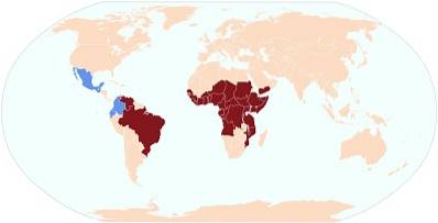
It is found mainly on the African continent, specifically in the sub-Saharan area, a site of which many parasites of the phylum Nematoda are endemic, such as Loa loa.
This parasite is found in the subcutaneous tissue of humans and is capable of triggering certain symptoms and signs that, together, constitute a pathology called onchocerciasis. It mainly affects the skin and eyes of infected people..
It is important to note that this parasite can live within its host for up to 10 years, causing serious damage and sequelae that greatly compromise their quality of life..
Article index
Onchocerca volvulus it is an organism grouped with multicellular eukaryotes, thanks to the fact that it has its genetic material packaged within the cell nucleus forming chromosomes. In addition, it is made up of different types of tissues, whose cells are specialized in various functions..
This nematode is triblastic, since during its embryonic development the three germ layers become evident: ectoderm, endoderm and mesoderm. The cells of these three layers differentiate and transform into different cell types to fulfill different functions, depending on the type of tissue they form. They are also deuterostomized.
This organism leads the life of a parasite, which is why, in order to develop, it needs to be inside the body of a host. Likewise, it is a pathogenic organism, since it is capable of causing an infection in humans known as onchocerciasis.
These parasites reproduce sexually, are ovoviviparous and present indirect development.
The taxonomic classification of Onchocerca volvulus is the next:
-Domain: Eukarya
-Animalia Kingdom
-Subkingdom: Eumetazoa
-Phylum: Nematoda
-Class: Secernentea
-Order: Spirurida
-Family: Onchocercidae
-Gender: Onchocerca
-Species: Onchocerca volvulus.
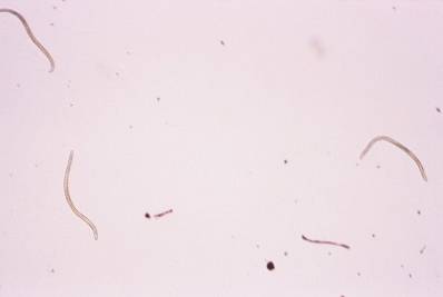
These nematode worms are cylindrical in shape and elongated in body. They have a kind of cuticle that covers their entire body. With regard to color, they are generally white.
They are dioecious, which means that the sexes are separated, that is, there are female individuals and male individuals..
Likewise, they present sexual dimorphism, which implies that there are certain aspects that allow differentiating females from males..
Because they present an indirect development, when they are born they do so in the form of larvae known as microfilariae. These are very small, barely reaching 300 microns and have a tail that is pointed..
They are much smaller than females. They are generally 5 cm in length. The terminal end of the body is curved. They also have two structures known as spicules that, in most cases, have different lengths..
In addition to this, compared to the female, the cuticle has a greater number of layers, in addition to having a wrinkled appearance.
The females are considerably larger than the males. They can reach up to more than 50 cm in length. Its rear end ends in a point, it is not curved like that of the males. Regarding the cuticle, the female has fewer layers and is not wrinkled in appearance, but rather has certain protrusions.
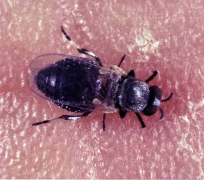
Onchocerca volvulus it is a parasite and as such requires a host as well as a vector to survive. The definitive host of this nematode is humans, while its vector is an insect, specifically a dipteran belonging to the genus Simulium.
Although there are several species within this genus, the one most frequently related to this parasite is Simulium damnosum.
This is in Africa, since, however, in the American continent, the species of this genus that are most related to this parasite are Simulium ochraceum, Simulium metallicrum Y Simulium callidum.
These insects are blood-sucking, that is, they feed on human blood by stinging.
The definitive host of this parasite is the human being. Inside, the microfilariae larvae (L1) are found mainly in the dermis. When the insect bites an infected individual, to the extent that it feeds on blood, it also ingests the microfilariae found on the skin.
Inside the animal's body, the parasite loses its protective covering (cuticle) and migrates from the stomach to the thoracic muscles. There it undergoes a metamorphosis, going from state L1 to L3. These L3 larvae migrate again towards the oral cavity of the insect, specifically to the proboscis.
When an infected fly bites a healthy human being, it transmits the L3 larvae, which enter the body through the wounds caused by the bite. In humans, they lodge in the subcutaneous tissue, where they undergo metamorphosis again, from L3 to L4 and L5, until finally reaching the adult state..
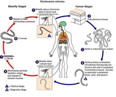
The adult worms remain in the subcutaneous tissue, usually in some connective tissue nodule. There they reproduce and the females begin to release microfilariae (L1 larvae). These larvae can be found on the skin for an average of 10-12 months after the parasite has entered humans..
The disease caused by the parasite Onchocerca volvulus it is known by the name of onchocerciasis. It is also known by other names such as river blindness, onchocerciasis and Robles disease, among other names..
Adult parasites cause a series of signs and symptoms to be triggered in the infected human being, at the level of various systems.
Some symptoms appear on the skin that are mainly related to irritation caused by the parasite.
The main symptom is pruritus (itching), edema (inflammation), as well as hyperthermia (increased temperature). Eventually, itching leads to skin irritation from excessive scratching.
Later, with the progress of the infection, hyperpigmented areas or areas that lose their pigmentation appear on the skin, as well as lesions called lichenified plaques..
If the infection is not treated, the skin loses its elasticity and a condition known as pachydermitis develops..
The presence of skin nodules, known as onchocercomas, is also common. These are located mainly at the level of the scalp.
One of the favorite tissues of these parasites in humans is the ocular conjunctiva. Due to this, infected people can present various symptoms at the ocular level.
Among the manifestations at eye level we can mention:
- This is sensitivity to light.
- Choroiditis: chronic inflammation of the choroid and retina.
- Uveitis: inflammation of the middle layer of the eye. Several structures such as the iris, choroid, and ciliary body are affected here..
- Sclerosing keratitis: it is an inflammation of the cornea. Here a permanent opacity of the cornea occurs.
- Optic nerve atrophy.
All these alterations greatly compromise the view. Eventually, a person with this infection loses vision to total blindness..
The progression of the infection can lead to neurological and kidney alterations. In addition to this, manifestations have been described at the level of the lymphatic system, such as obstruction of the lymphatic ducts. This obstruction leads to exaggerated inflammation. The hanging groin is a representative example of this..
The diagnosis of the disease is based both on the clinical observation of the symptoms and signs, as well as some tests that include a skin biopsy and a specialized eye examination..
If a doctor suspects that his patient may have the disease, he will proceed to take a skin sample (biopsy), which will be placed in saline for 24 hours and then proceed to observe it under a microscope. If microfilariae are seen, then it is positive for infection by Onchocerca volvulus.
Likewise, if the patient is suspected of having ocular involvement, he should undergo an examination using an instrument known as a slit lamp. This allows the doctor to visualize the eye in a magnified way and detect whether or not there are microfilariae or the adult parasite..
Blood tests are not reliable to accurately diagnose infection with Onchocerca volvulus, since there are other filarial-type parasites that can generate similar blood disorders.
Treatment of onchocerciasis is long-lasting. The medicine currently prescribed to treat this infection is an anthelmintic known as ivermectin. The form of administration is a single dose every six months. The duration time depends on the persistence of the symptoms.
The mechanism of action of this drug is based on the fact that it destroys microfilariae and, although it does not kill adult worms, it greatly reduces their fertility, such that they cannot produce microfilariae..
If the patient has onchocercomas, the doctor may make the decision to surgically remove them. Of course, the treatment is determined by the doctor's criteria, taking into account the severity and evolution of each particular case..


Yet No Comments