The panendoscopy, also known as upper digestive endoscopy (EDS), it is a technique established since the end of the last century, performed to observe the esophagus, stomach and the first portion of the small intestine, better known as the duodenum.
It was in 1868 that Kussmaul first introduced an open tube into the pharynx, using the light reflected by a gasoline lamp as illumination; A lot has changed since then. In its beginnings, this technique was merely diagnostic..
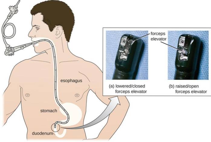
Over the years, science managed to implement instruments that allow treating certain procedures and obtaining tissues to analyze. This procedure is performed with the patient sedated and a flexible tube with a light and camera, called an endoscope, is inserted through the mouth without interfering with their breathing..
This tube will pass through the elements of the throat (pharynx and larynx), then the esophagus until it reaches the stomach and duodenum. It emits live images through a screen, which allows to perform pertinent interventions immediately, such as treating a small hemorrhage or taking a tissue sample for analysis (biopsy).
To be able to see better in the walls of the stomach, air will be introduced to distend it. The approximate time is 20 to 60 minutes. One of the problems with this test is that the patient, once finished, usually continues under the clouding effects of sedatives for a variable period of hours.
Article index
This procedure can be used in patients who come to a doctor's office presenting symptoms such as persistent pain in the upper abdomen, nausea, vomiting, swallowing problems or burning in the pit of the stomach..
It can even be symptoms that involve the voice and throat, such as dysphonia (hoarseness) or swallowing difficulties..
It can also be recommended when tumors, foreign bodies, bleeding in the upper part of the digestive tract, inflammation or ulcers in the esophagus, stomach or duodenum are suspected..
The suspicion of a tumor in the digestive tract is not limited exclusively to the patient alleging symptoms of discomfort, since by the time there are symptoms, the disease may already be at an advanced point in its natural history.
It is pertinent to identify the population that is at greater risk of presenting or developing cancer in any of the structures evaluated by this procedure, especially esophageal and stomach cancer, since they may not even present symptoms at any time.
Risk factors that justify performing this preventive or screening procedure (by detecting the disease in its early stages) include age, family history of cancer (especially stomach or esophagus), race (Asians are at higher risk of developing these tumors), and blood group (group A), among others.
Panendoscopy is often used to make a diagnosis. However, accessories can be attached to the endoscope for different purposes, such as removing foreign bodies (forceps), controlling areas of bleeding (alcohol, emboli), removing polyps or other superficial lesions..
It is also possible to take different tissue samples to analyze and thus detect tumors in the initial stages (biopsy), ultrasound techniques, and it can even be used to place crystals of radioactive material for the treatment of tumors; however, the latter is not a routine procedure (local radiotherapy).
Today the instruments used for panendoscopy have built-in ultrasound equipment that has specific uses, such as the diagnosis of infective endocarditis (infection of the inner walls of the heart), because just in front of the esophagus is the left atrium of the heart.
Another very important use of this tool is in esophageal cancer, since in early stages it tends to invade deep structures of the esophagus known as lymph nodes, a crucial step for the spread of the tumor in the body.
Before the test, the stomach must be completely empty. Therefore, the patient should not drink or eat anything in the 8 hours prior to the test..
You must inform if you suffer from heart or lung diseases, as well as give details of the medications you take and if you have any type of allergies.
This is important since the anesthetics used for the procedure can bring unfavorable reactions if the patient has an underlying disease or takes medications that interfere with the normal action of sedatives..
Endoscopy is the term used to describe the direct visual inspection of any part of the interior of the human body, through a flexible tube equipped with a mini-camera and guided by levers called an endoscope..
This instrument is introduced through natural orifices or through a minimal surgical incision. There are different types of endoscopy depending on the entrance orifice and the part of the body to be examined, these are:
Gastrocopy, as indicated by its etymology, refers exclusively to the visualization of the stomach, which can be through a natural or previously made orifice (for example, when the surface of the stomach is approached to feed patients with obstruction of the esophagus or throat).
Colonoscopy allows the colon or large intestine to be explored from the rectum to the lower end of the small intestine.
Bronchoscopy allows exploration of the trachea and bronchi. Like panendoscopy, the probe is inserted through the mouth.
Cystoscopy allows the urethra, bladder, and prostate to be seen in men. The endoscope is introduced through the urinary tract and covered with anesthetic gel.
It is a procedure that allows access to large joints (for example, knee). It has represented a great advance in sports medicine since its inception; thanks to this complex surgical procedures can be performed quickly and minimally invasive.
Panendoscopy is considered a very minimally invasive procedure and complications can include perforation or bleeding, reaction to the medication used for sedation, and infection of areas that have been cut or cauterized..
Doctors should always thoroughly discuss risks and complications with the patient before performing the procedure..


Yet No Comments