Papopavirus (Papovaviridae) is a family of small viruses that includes the Polyomavirus Y Papillomavirus. The organization of the genome between these viruses differs significantly. Therefore, some authors designate it as subfamilies, that is, subfamily Polyomaviridae and subfamily Papilomaviridae.
The Polyomaviridae contain JC virus isolated from brain tissues of patients with progressive multifocal leukoencephalopathy; BK virus, isolated from the urine of immunosuppressed kidney transplant recipients, causing hemorrhagic cystitis or nephropathy; and the SV40 virus, Simios 40 vacuolization virus that mainly affects these animals.
For their part, Papilomaviridae contain more than 70 serotypes of human wart virus, better known as Human Papillomavirus (HPV). These viruses are widely distributed worldwide.
These agents have a slow development cycle, stimulate cellular DNA synthesis, and replicate in the nucleus. Therefore, the infections they produce are latent and chronic in their natural hosts..
The suffering of these pathologies has been associated with the development of carcinogenic diseases in mammals.
In the case of papillomavirus, this occurs in natural hosts, where HPV infection is strongly related to the appearance of premalignant and malignant diseases of the vulva, cervix, penis and anus..
While in polyomaviruses the appearance of tumors has been observed only in experimental animals, with the exception of SV40, which produces tumors in humans..
Article index
These viruses have man and animals as their natural habitat. The form of transmission is by contact with infected secretions.
The routes of entry are cutaneous, genital (ETS) or respiratory for papillomaviruses, while for polyomaviruses it is unknown, but it is believed that it may be respiratory.
Both polyomaviruses and papillomaviruses, once they enter the body, remain latent in the tissues.
The pathologies can be treated, but if there is immunosuppression there may be relapses due to the reactivation of the virus.
HPV is divided into 2 groups according to its affinity for tissues: the cutaneous-tropics are those with a predilection for the skin, and the muco-tropics are those with the highest affinity for mucosa.
Among HPV serotypes, associations have been seen between certain genotypes and the type of clinical lesion. There are also serotypes more oncogenic than others. For example, the HPV 16 and HPV 18 serotypes that cause genital condylomata are high risk.
In the case of the HPV-16 serotype, it is associated with keratinizing squamous cell carcinomas, while HPV-18 is associated with adenocarcinomas.
Likewise, in patients affected by verruciform epidermodysplacia by HPV serotypes 5 and 8, a high rate of subsequent suffering of squamous cell carcinoma from the lesions is recorded..
In summary, the high-risk serotypes are: 16, 18, 31, 33, 35, 39, 45, 51, 52, 56, 58, 59, 68, 82, 26, 53, 66. And low-risk: the 6, 11, 40, 42, 43, 44, 54, 62, 72, 81.
DsDNA Group 1.
Family: Papovaviridae.
Genus: Polyomavirus and Papillomavirus.
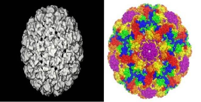
The Papovavirus in general, they are 45-55 nm in size, icosahedral symmetry, and do not have a lipid envelope. They possess a circular double-stranded DNA genome.
Polyomaviruses consist of two or 3 replicative genes called tumor antigens encoded by one of the DNA strands and three structural genes, called capsid antigens encoded on the other strand..
Human and animal polyomaviruses are antigenically distinct, with only one serotype of each. The prototype virus is the Ape virus 40 of monkeys.
Papillomaviruses are similar to polyomaviruses, however they present certain differences. Among them: viral particles have a diameter of 55 nm and the structure of the genome is more complex. All viral genes are encoded on a single strand of DNA.
The HPV virus contains 2 proteins L1 and L2, and also has viral oncoproteins that interact with cell tumor suppressor proteins.
In humans, they produce latent infections at various sites depending on the virus. For example, KV and SV40 viruses persist in kidney cells.
While the JC virus remains latent in the tonsillar tissue, in the stromal tissue of the bone marrow, in the epithelial cells of the colon and kidney, among other tissues, indefinitely..
Most infections are asymptomatic. These viruses reactivate and cause symptomatic disease only in immunosuppressed patients..
In HPV, the scales from the exfoliation of the skin are an important source of contagion, as is sexual contact.
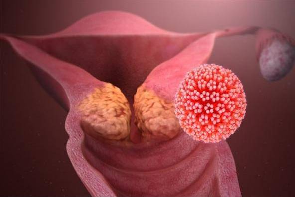
The human papillomavirus has a predilection for infecting cells of the attachment site of the squamous and columnar epithelium, the vulva, cervix and anus being the most vulnerable sites.
The replication and assembly of the virus occurs in the layers of the squamous epithelium in the process of differentiation, since the virus initially infects the basal layer of the epithelium, where the viral DNA is located..
But the expression of the capsid proteins and the assembly of the complete virus occurs in the most superficial layer of differentiated keratinocytes, that is, when the cells finish their maturation.
Therefore, to be able to replicate, the virus needs the cells to be in the process of differentiation (maturation), and due to this it has not been possible to be cultivated in vitro, because although there are cell cultures, they cannot complete their differentiation stage under these conditions. and therefore the virus cannot replicate either.
It should be noted that the HPV virus can establish a lytic infection in the keratinized cells of the superficial epithelium or it can remain dormant in the deeper layers, persisting for years in it..
Likewise, it is important to note that the cells that shed or shed from the affected epithelium will be loaded with virus, helping its dissemination..
On the other hand, if DNA is integrated into cellular DNA, it can cause an oncogenic transformation of the host cell..
In this way, the viral genes E6 and E7 are activated, causing damage to the p53 gene of the basal cell. This gene is responsible for correcting errors that can occur during cell reproduction. When the gene is damaged it cannot exert its function, therefore the cells become neoplastic.
On the other hand, the virus produces an oncogenic protein p105 and forms a complex with the RB gene to damage it..
The RB gene controls and regulates cell reproduction, telling cells when to reproduce and when to stay at rest.
By blocking its function, cells reproduce without stopping and become cancerous.
The JC virus is neurotropic and causes progressive multifocal leukoencephalopathy. This rare disease attacks immunosuppressed patients. The virus replicates in oligodendrocytes producing a demyelination of the central nervous system (destructive encephalitis).
Likewise, the virus stimulates the immune system and induces a humoral and cellular immune response (cytotoxic T), controlling the infection that remains latent. The virus is reactivated when the immune system is depressed, being the deterioration of cellular immunity essential for the development of the disease.
Interferon can inhibit polyomavirus, although it is weakly induced during infection.
The JC virus causes tumors in laboratory mice, but not in humans. Both the JC, BK and SV40 viruses have been associated with cases of hemorrhagic cystitis and progressive multifocal leukoencephalopathy.
While, BK and SV40 are also associated with cases of nephropathy.
On the other hand, SV40 has been associated with some tumors in humans, including primary brain tumors, malignant mesotheliomas, bone cancers, and non-Hodgkin's lymphomas..
Regarding the form of transmission of the JC and BK viruses, it is unknown, but it is believed that it may be through the respiratory route, while the vacuolizing simian virus 40 has affected humans due to the accidental contamination of polio vaccines with SV 40 virus.
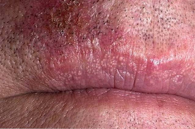
Papillomaviruses are responsible for benign papillomatous lesions of the skin and mucous membranes.
These lesions can present as common warts, flat warts, plantar warts, anogenital warts, epidermodysplasia verruciform, and laryngeal papillomas..
On the other hand, there is a very close association between the appearance of cervical intraepithelial neoplasia, cervical cancer, and respiratory tract tumors with human papillomavirus infection..
A simple test for the prevention of cervical cancer is the annual endocervical cytology test, stained with the papanicolaou technique. This test reveals pathognomonic features of HPV infection..
The diagnostic characteristic of the cell infected with HPV is koilocytosis, that is, the presence of a perinuclear halo of the squamous epithelium accompanied by nuclear atypia.
Molecular biology tests are necessary to identify the serotype involved. Likewise, colposcopy is a technique that helps to look for lesions on the cervix that may be caused by HPV..
VBK DNA can be detected in urinary sediment, in blood or in cells infected with viral inclusions, from kidney or urothelial tissue samples, through a PCR DNA detection study..
For the diagnosis of JC virus progressive multifocal leukoencephalopathy, the clinical aspect is important and the use of imaging and laboratory studies is also helpful..


Yet No Comments