
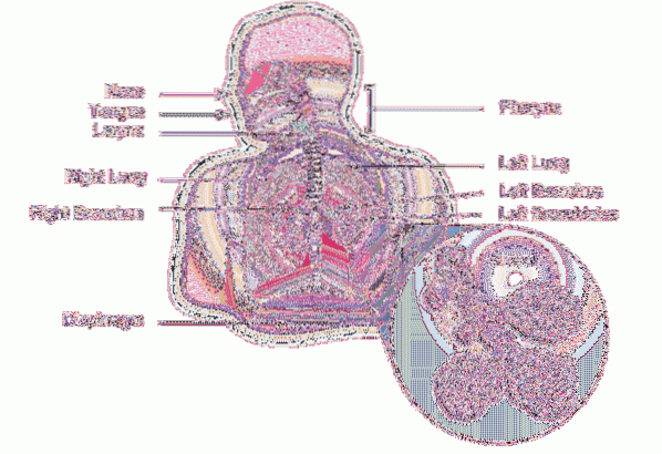
The Pulmonary parenchyma It is the functional tissue of the lung. It is made up of an air conduction system and a gas exchange system. It has different structural components in the tubes and ducts that constitute it from the nose to the pulmonary alveoli..
Around the pipe system, the lung parenchyma has elastic and collagen fibers arranged in the form of a mesh or network that has elastic properties. Some elements of the piping system have smooth muscle in their structure, which allows the diameter of each tube to be regulated..
The lung does not have muscles that allow its expansion or retraction, this function is fulfilled by the muscles of the rib cage, which are called "respiratory muscles". The lungs, from this point of view, are organs that passively follow the movements of the “box” that surrounds them..
There is also no ligament or structure that fixes the lungs to the rib cage, both hang from their respective main bronchi, the right bronchus and the left bronchus, and both the rib cage and the lung are covered with a membrane called the pleura..
Diseases of the lung parenchyma can be simply classified as infectious diseases, tumor diseases, restrictive diseases, and obstructive diseases..
An environment free of toxic substances and smoke or suspended particles and not consuming drugs by inhalation or cigarettes prevents many of the main diseases that affect the lung parenchyma and, therefore, respiratory function.
Article index
The lungs are two organs located within the rib cage. They are composed of a system of pipes that undergoes 22 divisions called “bronchial generations”, which are found before reaching the alveolar sacs (23) which are the gas exchange sites where the respiratory function is fulfilled..
From the main bronchi to the 16th bronchial generation, the airways fulfill exclusively conduction functions. As the routes are subdivided, the diameter of each tube in particular becomes smaller and smaller and its wall increasingly thin.
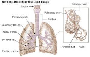
When the walls of the pipe system lose cartilage, its name changes from bronchus to bronchiole, and the last generation of bronchial tubes with exclusive conduction function is called terminal bronchiole..
Starting from the terminal bronchiole, the following bronchial generations are called respiratory bronchioles, until giving rise to the alveolar ducts and ending in the alveolar sacs or alveoli..
The only function of the alveoli is the exchange of gases (O2 and CO2) between the alveolar air and the blood that circulates through the alveolar capillaries and forms a network or capillary mesh around each alveolus..
This structural subdivision of the airways makes it possible to increase the surface available for gas exchange. If each of the alveoli is extracted from one lung, stretched and placed side by side, the surface area reaches between 80 and 100 m2, which is more or less the surface of an apartment.
The blood volume in contact with this enormous surface area is approximately 400 ml, which allows the red blood cells, which are the ones that carry O2, pass one after the other through the pulmonary capillaries..
This huge surface area and an extremely thin barrier between the two gas exchange territories provide the ideal conditions for this exchange to take place quickly and efficiently..
The lung and rib cage are attached to each other through the pleura. The pleura is made up of a double membrane made up of:
- A leaf that receives the name of leaf or parietal pleura, which is strongly adhered to the internal surface of the rib cage covering its entire surface.
- A sheet called the visceral pleura, tightly attached to the outer surface of both lungs.
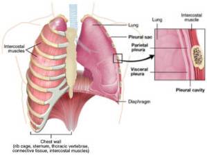
Between the visceral and parietal leaf there is a thin layer of liquid that allows the two leaves to slide relative to each other, but which generates great resistance for the separation of both leaves. For this reason, the visceral and parietal leaves of the pleura are held together and thus the thoracic wall and the lung remain together..
When the chest wall expands as a result of the respiratory muscles, the lung follows, through its pleural junction, the movements of the cage and, therefore, is distended, increasing its volume. When the anterior muscles relax, the box retracts, reducing the size of each lung..
From the first breaths that occur at birth, both lungs expand and acquire the size of the rib cage, establishing the pleural relationship. If the rib cage opens or air, blood or fluid enters the pleural cavity in a significant way, the pleurae separate.
In this case, the lung whose parenchyma has abundant elastic tissue and which was expanded or stretched due to the effect of the pleural relationship, now retracts (as a stretched elastic band does) loses all the air and remains hanging from its main bronchus.
When this happens, the rib cage expands, becoming larger than it was when it was attached to the lung. In other words, both organs acquire their independent elastic rest position.
The intrapulmonary conduction system is made up of the different bronchial divisions starting from the secondary or lobar bronchi. The bronchi have a respiratory epithelium that is pseudostratified and is made up of basal cells, goblet cells, and ciliated columnar cells..
The bronchial wall is covered with sheets of cartilage that give it a rigid structure that offers resistance to external compression, so the bronchi tend to remain open. Elastic and smooth muscle fibers are found around the tube in a helical arrangement..
The bronchioles do not have cartilage, so they are subjected to the traction forces exerted by the elastic tissue that surrounds them when it is stretched. They offer very little resistance to all the external compressive forces that are applied to them, therefore, they can easily and passively change their diameter..
The epithelial lining of the bronchioles varies from a simple ciliated epithelium with scattered goblet cells (in the larger ones), to a ciliated cuboid epithelium without goblet cells and clear cells (in the smaller ones).
Clear cells that are cylindrical cells with a dome-shaped top or apex and short microvilli. They secrete glycoproteins that cover and protect the bronchial epithelium.
The alveoli are about 300,000,000 in total. They are arranged in bags with many partitions; They have two types of cells called type I and type II pneumocytes. These pneumocytes are attached to each other through occluding junctions that prevent the passage of fluid.
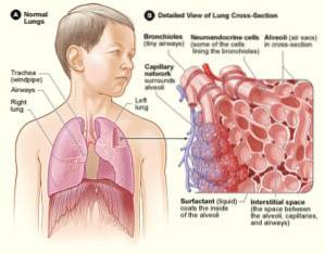
Type II pneumocytes are more prominent cuboid cells than type I. In their cytoplasm they contain laminar bodies and these pneumocytes are responsible for synthesizing the pulmonary tensioactive substance that covers the internal surface of the alveolus and lowers surface tension..
The alveolar and endothelial basal laminae fuse and the thickness of the alveolar-capillary barrier that gases must pass through to pass from one side to the other is minimal.
The tissue that surrounds the piping system has a hexagonal arrangement, it is made up of elastic fibers and collagen fibers that are rigid. Its geometric arrangement forms a net, similar to a nylon stocking, which is made up of rigid individual fibers woven into an elastic structure.
This conformation of elastic tissue and elastic interlocking structure gives the lung its own characteristics, which allow it to passively retract and, under certain expansion conditions, offer minimal resistance to distension..
Pulmonary diseases can be of infectious origin due to bacteria, viruses or parasites that affect the lung tissue.
Tumors of a different nature, benign or malignant, can also form, capable of destroying the lung and causing the death of the patient due to lung or brain problems, which are the most important areas of lung metastasis..
However, many diseases of various origins can cause obstructive or restrictive syndromes. Obstructive syndromes cause difficulty for the entry and / or exit of air from the lung. Restrictive syndromes cause respiratory distress by reducing the lung's ability to expand.
Examples of obstructive diseases include bronchial asthma and pulmonary emphysema..
In bronchial asthma, the obstruction is due to an active, allergic contraction of the bronchial musculature.
Contraction of the bronchial muscle reduces the diameter of the bronchi and makes it difficult for air to pass. Initially the difficulty is greater during expiration (air out of the lung) since all the retraction forces tend to close the airways even more.
In the case of pulmonary emphysema, what occurs is a destruction of the alveolar septa with loss of elastic lung tissue or, in the case of physiological emphysema in adults, the interwoven structure of the lung parenchyma is altered..
In emphysema, the decrease in elastic tissue decreases the pulmonary retraction forces. For any lung volume that is examined, the diameter of the pathways is reduced as external elastic traction is reduced. The end effect is respiratory distress and air trapping..
Lung restrictive syndrome is due to the replacement of elastic tissue by fibrous tissue. This reduces the lung's ability to stretch and causes shortness of breath. These patients breathe with smaller and smaller volumes and higher and higher respiratory rates..


Yet No Comments