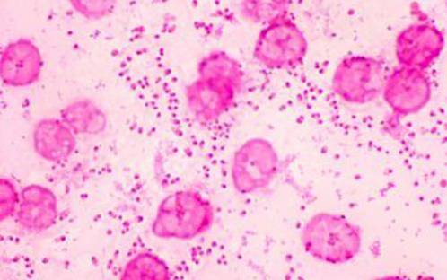
Pasteurella multocida characteristics, morphology, pathogenesis
Pasteurella multocida is a non-mobile gram-negative bacterium belonging to the Pasteurellaceae family, which are normally found in the flora of the upper respiratory tract and gastrointestinal tract of some species of animals, such as cats, dogs, pigs, rabbits, among others.
In 1879, the French veterinarian Henri Toussaint succeeded in isolating for the first time the Pasteurella multocida, while researching cholera disease in chickens. Since that time, this bacterium is considered one of the main causative agents of several infections in man and in animals, both wild and domestic..

Among the conditions caused by this bacterium are hemorrhagic septicemia and pneumonic pasteurellosis in cattle, atrophic rhinitis in pigs, rhinopneumonitis in rabbits, and cholera in chickens..
In man it could lead to affections at the level of the nervous, cardiovascular, respiratory system, among others..
Article index
- 1 Vaccine
- 2 Features
- 2.1 Transmission modes
- 2.2 Carriers
- 2.3 Epidemiology
- 2.4 Microscopic
- 2.5 Capsules
- 2.6 Metabolic properties
- 3 Taxonomy
- 3.1 Subspecies of Pasteurella multocida
- 4 Morphology
- 4.1 Shape and size
- 4.2 Movement
- 5 Pathogenesis
- 5.1 -Symptomatology of infection in humans
- 5.2 -Symptoms of infection in animals
- 6 Treatment in humans
- 7 References
Vaccine
The chemist and bacteriologist Louis Pasteur carried out, in 1880, some experiments to know the mechanism of transmission of the Pasteurella multocida, since at that time it was causing the death of many poultry. The work consisted of inoculating the bacteria in healthy chickens to evaluate the disease.
As a result of his research, he observed that the bacteria could be weakened, to the point that when injected into birds they made them immune to the disease.
This is how he discovered that it was not necessary to find a specific bacterium to vaccinate the animals, the P. multocida bacteria themselves could be weakened and used as vaccines.
Characteristics
Transmission modes
In a high percentage, humans are directly infected if they are bitten or scratched by a cat or dog that has the bacteria. To a lesser extent, cases of infection due to the bite of rodents or rabbits have been reported..
The bacteria could also be transmitted indirectly through contact with secretions such as saliva or excretions of infected animals. There is no documentation of transmission between two people or by consumption of contaminated food or water.
Carriers
Some of the animals that can be carriers, and suffer from the diseases that this bacterium produces, can be rabbits, pigs, cows, cats, dogs, chickens and turkeys.
epidemiology
The Pasteurella multocida It is located in the digestive system, especially in the gastrointestinal tract, and in the upper respiratory tract of mammals and poultry, which are the main reservoirs of this bacterium.
Some epidemiological studies indicate that only 3% of humans who have been in contact with infected animals have been infected by P. multocida strains.
This percentage increases if the person has a history of respiratory disease, if they are older than 60 years or if they suffer from some type of immunosuppressive disease..
Microscopic
These bacteria do not stain deep blue or violet on Gram's stain. Rather, they take on a faint pinkish coloration..
Capsules
The ability of this bacterium to invade and reproduce in the host increases thanks to the presence of a capsule formed by polysaccharides that surrounds it. This is because it allows it to easily evade the innate response of the P. multocida host..
It can be classified into five different groups (A, B, D, E and F), which have different chemical compositions. In type A strains, the capsule is mainly made up of hyaluronic acid. It is associated with fowl cholera, rhinopneumonitis in rabbits and respiratory problems in ruminants, pigs, dogs and cats..
Type B contains galactose, mannose, and the polysaccharide arabinose. They are present in the bacteria responsible for hemorrhagic septicemia in cows. Those of type D have heparin, being related to atrophic rhinitis in pigs and pneumonia in ruminants.
Regarding the type E, there is still no clear data on their biochemical structure, however, it is presumed that they are part of the bacterium that causes septicemia in cattle. In P. multocida of capsular type F, the constitution is made up of chondroitin and is related to cholera in turkeys.
Metabolic properties
They are facultative anaerobic, requiring a PH between 7.2 and 7.8 to reach their development. They are chemoorganotrophic, since they obtain energy as a product of the oxidation of some organic compounds. Metabolisms can be fermentative or respiratory.
This bacterium can be differentiated from other species due to its absence of hemolysis in environments where blood is present, the production of indole and the negative reaction to urea.
Taxonomy
Kingdom: Bacteria.
Subkingdom: Negibacteria.
Phylum: Proteobacteria.
Class: Gammaproteobacteria.
Order: Pasteurellales.
Family: Pasteurellaceae.
Genus: Pasteurella.
Species: Pasteurella aerogenes, Pasteurella bettyae, Pasteurella caballi, Pasteurella canis, Pasteurella dagmatis, Pasteurella langaaensis, Pasteurella lymphangitidis, Pasteurella mairii, Pasteurella multocida, Pasteurella oralis, Pasteurella pneumotropica, Pasteurella skyensis, Pasteurella stomatis, Pasteurella testudinis.
Subspecies of the Pasteurella multocida
Pasteurella multocida gallicida
This is recognized as the main causal agent of cholera in birds, although it has also been identified in cattle. Its biochemistry shows that it contains sucrose, dulcitol, mannitol, sorbitol and arabinose.
Pasteurella multocida multocida
It has been found in cattle, rabbits, dogs, birds, pigs, and chickens. The species causes pneumonia in ruminants and pigs, and avian pasteurellosis or cholera in chicken, turkey, ducks and geese. Biochemically contains sucrose, mannitol, sorbitol, trehalose and xolose.
Septic pasteurella multocida
It has been isolated in different species of felines, birds, canines and in humans. It is made up of sucrose, mannitol and trehalose.
Morphology
Shape and size
They are coccoids or cocobacillaries, which implies that they could have a short rod shape, intermediate between cocci and bacilli..
They have pleomorphic cells with a rod-like shape, which can appear individually in groups of two or in short chains, convex, smooth and translucent. Its size can range between 0.3-1.0 by 1.0-2.0 micrometers.
Movement
The Pasteurella multocida it is an immobile bacterium, so it does not have flagella that allow it to move.
Pathogeny
The bacteria Pasteurella multocida it is usually a commensal in the upper respiratory tract of some domestic and wild animals. Infection in humans is associated with biting, scratching or licking.
Initially, the infection presents with inflammation of the deep soft tissues, which can manifest as tenosynovitis and osteomyelitis. If these become severe, endocarditis may develop.
-Symptoms of infection in humans
Local
There may be redness, pain, tenderness and some purulent-type discharge. If it is not treated in time, an abscess could form in the area.
Respiratory system
Hoarseness, sinus tenderness, pneumonia, and redness of the pharynx may occur.
Central Nervous System
Clinical cases have been reported in which, possibly due to infection by P. multocida, there is some focal neurological deficit or stiff neck.
Ocular
An ulcer may appear on the cornea, which results in a decrease in the visual acuity of the infected person..
Circulatory system
Hypotension and tachycardia could be symptoms of infection by Pasteurella multocida, as well as inflammation of the pericardium, the membrane that covers the heart.
Reproductive system
On rare occasions, there have been cases where men may present an inflammation of the epididymis, while in women the cervix may present cervicitis.
Excretory system
The excretory system may be affected with pyelonephritis, an inflammation of the kidney that can cause groin pain and fever..
-Symptoms of infection in animals
Animals infected with the bacteria may present asymptomatic or mild infections in the upper respiratory organs. In this case they could suffer from pneumonia, with fatal consequences for the animal.
Some symptoms could be rhinitis, with sneezing accompanied by mucous secretions and fever. Transmission between animals occurs by direct contact with nasal secretions.
Treatment in humans
The treatment of this infection is usually based on the use of penicillin, since the different species of Pasteurella multocida they are organisms very sensitive to this type of antibiotic.
References
- ITIS (2018). Pasteurella. Recovered from itis.gov.
- Wikipedia (2018). Pasteurella multocida. Recovered from en.wikipedia.org
- Sara L Cross, MD (2018). Pasteurella Multocida Infection. Medscape. Recovered from emedicine.medscape.com.
- John Zurlo (2018). Pasteurella species. Infectus disease advisor. Recovered from infectiousdiseaseadvisor.com.
- Clinical Veterinary Advisor (2013). Pasteurella multocida. ScienceDirect. Recovered from sciencedirect.com.
- Stephanie B. James (2012). Children's Zoo Medicine. ScienceDirect. Recovered from sciencedirect.com.
- Yosef Huberman, Horacio Terzolo (2015). Pasteurella multocida and Avian Cholera. Journal of Veterinary Medicine Argentina. Recovered from researchgate.net.
- David DeLong (2012). Bacterial Diseases. SicenceDirect. Recovered from sciencedirect.com.
- Veterinary bacteriology. Swiss University of Agriculture (2018). Pasteurella multocida subsp. multocida. Recovered from vetbact.org.
- Fiona J. Cooke, Mary P.E. Slack (2017). Gram-Negative Coccobacilli. ScienceDirect. Recovered from sciencedirect.com.


Yet No Comments