
The brain peduncles They are brain casts made up entirely of nerves. Each human brain has two cerebral peduncles that are joined by an interpeduncular fossa.
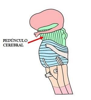
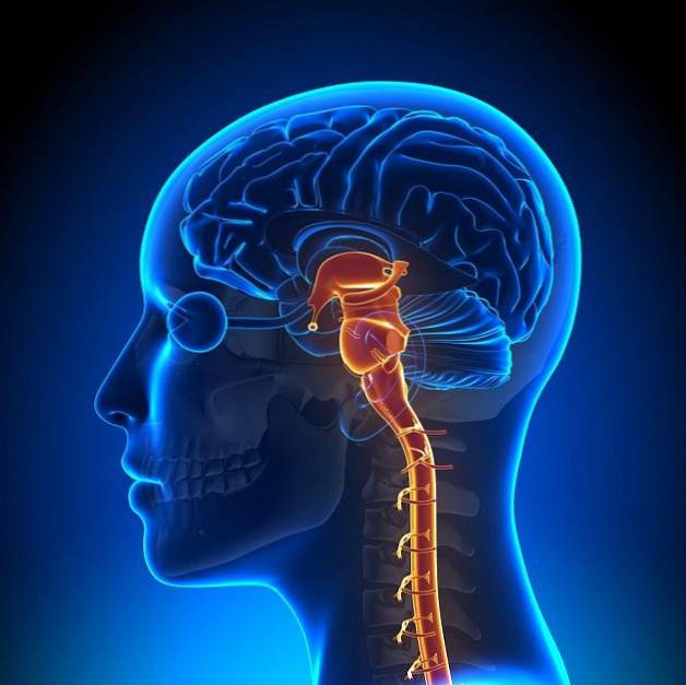
The cerebral peduncles are located in the upper region of the brainstem, just above the annular pons. Very extensive brain regions result that extend throughout the entire length of the brain until reaching the cortex. In the left and right hemispheres of the cerebral cortex, the cerebral peduncles disappear.
The cerebral peduncles are important structures that are in charge of joining and communicating the midbrain with the brain. In this sense, these structures perform functions related to the reflex control of movements.
Article index
The cerebral peduncles are two masses or nerve cords; They have a cylindrical shape and are white. Both cerebral peduncles are separated from each other by an interpeduncular fossa or posterior perforated space.
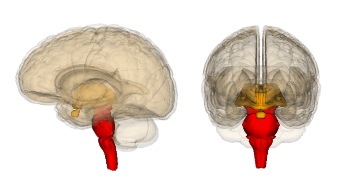
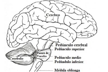
They are located in the upper part of the brainstem, that is, the brain region composed of the midbrain, the bridge of Varolio and the medulla oblongata.
Specifically, the cerebral peduncles are just above the bridge of Varolio. However, its structure is longer than that of the other regions of the brain stem, extending to the cerebral hemispheres.
The cerebral peduncles are also known as the basis pedunculi and are found in their entirety (except the tectum) within the midbrain.
The main function of these brain regions is to communicate the midbrain with the brain. They are involved in the reflex control of eye movements and in the coordination of these movements with the head and neck.
The three brain regions that give rise to the cerebral peduncles are the cortex, the spinal cord, and the cerebellum..
The cerebral peduncles include the tegmentum of the midbrain, the cerebral crus and the pretectum, and present numerous nerve pathways that inside.
Specifically, in the peduncular cerebral circuit, the fibers of the motor areas of the brain project to the cerebral peduncle and, later, project to different thalamic nuclei.
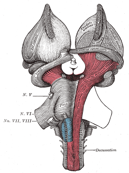
Anatomically, the cerebral peduncles are structured by nerve fibers, which include the fibers of the corticopontine tracts (which is in charge of communicating the cerebral cortex with the Varolio bridge) and the corticospinal tract (which faces the union of the cerebral cortex with the spinal cord).
Regarding its structure, in the cross section each peduncle has a dorsal region and a ventral region, which are separated by a layer of pigmentation of the gray matter (the black substance)..
In this sense, the two main parts of the cerebral peduncles are: the cerebral crus and the tegmentum..
The cerebral crus is the front part of the cerebral peduncle. It is an extension of nerves in the shape of a leg that transmits brain impulses to the relevant regions of the body to control movement.
The information that emerges from the cerebral cross of the peduncles is the result of the interaction between the conscious decision to move that is carried out in the cerebral cortex, and the modifications made in the brainstem through the information received about the position and current state of body.
The cerebral cross of the peduncles receives complete information about the movements to be transmitted to the organism, taking into account both the planning of the movement and its adaptation to the real circumstances of the body..
The tegmentum or covering is the posterior region of the cerebral peduncles. It is a structure that presents a very early embryonic development and constitutes a basic region for communication between the cortex and the brain stem..
The tegmentum of the cerebral peduncles is characterized by sending and receiving information from both the cerebral cortex and the brain stem.
This action of the peduncle allows the development of refined information that is transmitted directly to the cerebral crus, that is, to the other region of the peduncle..
When the tegmentum of the cerebral peduncles is damaged, the body changes its movement pattern. The person is unable to carry out natural actions and acquires a robotic movement.
The cerebral peduncles have two main functions: the conduction of impulses and the development of reflex acts..
Regarding impulse conduction, the cerebral peduncles are basic structures that allow the midbrain to be connected to the brain.
The brain is a structure that includes the cerebral cortex, the telencephalon, and the diencephalon. These brain regions contain important structures that allow the development of most brain activities.
However, for many of the actions carried out by these structures to be carried out, it is necessary that they be transmitted to lower regions and, in some cases, to the spinal cord and specific body regions.
In this sense, the cerebral peduncles allow the transmission of information from the brain to the midbrain (and vice versa).
When information comes from lower structures, the brain peduncles collect information from the midbrain to carry it to the brain. On the other hand, when the nerve impulses come from higher structures, it is the cerebral peduncles themselves who are responsible for transmitting the information to the midbrain..
Regarding reflex movements, cerebral peduncles are characterized by intervening in the control of eye movements and the coordination of these movements with the head and neck.
It is important to emphasize that the cerebral peduncles are not the same structures as the cerebellar peduncles..
In this sense, the cerebellar peduncles would be structures comparable to the cerebral peduncles pertinent to the cerebellum..
In this case, the cerebellar peduncles seem to perform functions of integration of the information received, with the aim of controlling the orders that the cerebral cortex sends to the locomotor system..


Yet No Comments