Pepto-streptococcus It is a genus of bacteria formed by species of Gram positive anaerobic cocci of variable size and shape. They are found as part of the normal microbiota of mucous membranes, especially oropharyngeal, intestinal and genitourinary.
They are a frequent cause of mixed or polymicrobial infections of endogenous origin. They can be isolated from cultures of brain and liver abscesses, bacteremia, pleuropulmonary infections, vulvar, Tubovaric and pelvic abscesses, among others..
Among its main species are P. anaerobius, P. asaccharolyticus, P. indolicus, P. magnus, P. micros, P. prevotii, P. productus and P. tetradius. Other lesser known are P. hydrogenalis, P. ivorii, P. lacrimales, P. lactolyticus, P. octavius, P. vaginalis, among others.
Article index
Species of the genus Peptoestreptococcus are obligate anaerobes, that is, they do not grow in the presence of oxygen. They do not form spores and are non-motile.
Many of the species are part of the normal human microbiota and are harmless as long as they remain in healthy mucosa. But they are opportunistic pathogens as they enter the deep tissues near these areas..
That is why the species of the genus Peptoestreptococcus have been involved in some infectious processes. For example: Peptoestreptococcus anaerobius has been isolated from clinical specimens of the mouth, upper respiratory tract, skin, soft tissues, bones, joints, gastrointestinal and genitourinary tract. P. stomatis has been isolated from the oral cavity.
Although not much is known, it is known that certain strains of Peptoestreptococcus possess electron microscopic demonstrable capsule and some oral strains produce hyaluronidase.
Both the presence of the capsule and the production of hyaluronidase represent virulence factors. Likewise, the content of fatty acids in the cell wall of certain strains of Peptoestreptococcus is characteristic, but its participation as a virulence factor is unknown..
On the other hand, it must be taken into account that infections caused by anaerobic bacteria are generally polymicrobial, with a synergism between the different species..
This means that the various bacteria that make up the mixed infection share, so to speak, their virulence factors with each other, which compensates for the lack of pathogenicity factors of certain strains..
For example, the presence of Bacteroides will provide Betalactamases that will protect Pepto-streptococcus that are sensitive to penicillins..
Likewise, other facultative bacteria will use the oxygen that may be present, which produces a more suitable medium for strict anaerobes such as Pepto-streptococcus..
Domain: Bacteria
Phylum: Firmicutes
Class: Clostridia
Order: Clostridiales
Family: Peptoestreptococcaceae
Genus: Peptoestreptococcus
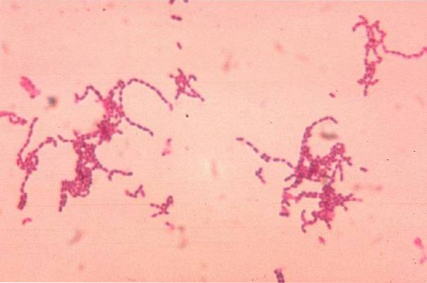
Pepto-streptococci seen under a Gram-stained light microscope are Gram-positive cocci and some species may appear coccobacillary and form chains. In old cultures they are usually Gram negative.
There are some differences in the appearance and distribution of microorganisms depending on the species. Among them the following can be highlighted:
Peptoestreptococcus anaerobius Y P. products are large coccobacilli that often form chains.
Instead P. magnus It is more coccoid, measures> 0.6 μm in diameter and occurs isolated or in mass similar to the Staphylococcus sp.
Peptoestreptococcus micros measure < de 0,6 μm de diámetro y se presenta formando cadenas cortas. En tanto que, P. tetradius presents as unusually large coconuts in clumps.
The development of these anaerobic cocci in broth is typically slow and forms spheres, clumps, or aggregates rather than diffuse turbidity..
They form tiny, convex, gray to white, opaque colonies. Its edges are whole; the surface may appear "pitted" or marked by depressions.
The size of the colony ranges from 0.5-2 mm in diameter and a halo of discoloration can be observed around it (P. micros).
On special blood agar for anaerobes P. micros may cause slight beta hemolysis.
In infection with Pepto-streptococcus, the breakdown of an anatomical barrier (mucosal surface, skin) plays a fundamental role, which leads to the introduction of these bacteria to normally sterile sites..
There are sites that are more susceptible to creating hypoxic conditions due to the presence of facultative microorganisms that help reduce oxygen and limit the local potential for redox, favoring anaerobic infections..
These sites are the sebaceous glands of the skin, the gingival clefts of the gums, the lymphoid tissue of the throat, and the lumen of the intestinal and urogenital tracts..
On the other hand, it is common to observe these infections in immunosuppressed patients, where most infections occur with mixed flora (polymicrobial), almost always of endogenous origin..
The characteristics of infections caused by Pepto-streptococcus are not very different from those of other anaerobic bacteria. These characteristics are as follows:
Brain abscesses They occur by extension through the lamina cribrosa of the ethmoid into the temporal lobe, which gives the typical location of these abscesses.
They have been involved in periodontal infections, otitis, etc..
Especially Pepto-streptococcus micros is a recognized pathogen in dental infections (progressive periodontitis), where Chlorhexidine does not eradicate the microorganism.
Likewise, P. vaginalis has been isolated from the conjunctival mucosa and ears.
It can be caused by human bites.
Necrotizing pneumonia, lung abscess. Occur due to aspiration of oropharyngeal content.
Peritonitis, cholangitis, abscesses. They originate from the eruption of the intestinal mucosa.
Tubo-ovarian abscesses, pelviperitonitis, septic abortions, endometritis, pelvic inflammatory disease.
They have been isolated from cervical epidural abscess and cerebrospinal fluid. This is possible due to contamination during previous surgical procedures..
Non-clostridial anaerobic cellulitis, necrotizing fasciitis.
It must be carried out by qualified personnel, since the sample collection and transfer must be carried out with extreme care, avoiding exposure to oxygen..
The most commonly used transport medium is Stuart, which consists of a buffer solution of sodium and potassium chloride, magnesium and potassium chloride, thioglycollate and agar..
The buffer helps maintain the proper pH so that the microorganism remains viable. Thioglycollate is added as a reducing agent to enhance the recovery of anaerobic bacteria.
While agar provides a semi-solid consistency to the medium to prevent oxygenation and spillage of the sample during transport.
Sowing is done in special media for anaerobes. For example, Blood Agar is prepared from soybean trypticase with 5% sheep's blood..
In some cases it is supplemented with yeast extract, hemin, vitamin K or L-cystine for demanding anaerobes.
Phenylethyl alcohol can also be added to inhibit the growth of enterobacteria or antibiotics such as kanamycin and vancomycin, among other formulations to inhibit facultative anaerobic Gram negative bacilli..
On the other hand, cultures in liquid media such as enriched thioglycollate and glucose-minced meat should be maintained for a minimum of 5 to 7 days before discarding them as negative..
The seeded plates should be immediately placed in anaerobic jars with a commercial envelope (GasPak).
This envelope catalytically reduces oxygen by hydrogen generated along with carbon dioxide. Under this anaerobic environment, the plates are incubated for at least 48 hours at an optimal temperature of 35ºC to 37ºC..
Exposure of freshly seeded plates to ambient oxygen for 2 hours can inhibit or delay the growth of this genus, so they should be seeded and incubated immediately..
It should be borne in mind that in cases of bacteremia due to Peptoestreptococcus anaerobius the addition of Sodium Polyanethole Sulfonate (SPS) to blood culture bottles inhibits the proliferation of this microorganism.
This same substance placed in the shape of a disc on the crop seed serves to differentiate Peptoestreptococcus anaerobius of other Pepto-streptococcus species, by observing a halo of inhibition around the disc.
The genus Peptoestreptococcus is considerably susceptible to most antibiotics, although it is especially resistant to tetracycline, erythromycin, and occasionally to cefamandeol and ceftazimide..
Some strains that previously belonged to the genus Peptococcus and were later transferred to the genus Pepto-streptococcus cannot be treated with clindamycin..
In summary, treatment should include debridement, drainage and cleaning of the affected area, use of antimicrobials, and placement of hypervaric oxygen. The use of antibiotics alone will not solve the problem, due to the inability to penetrate the site of infection.
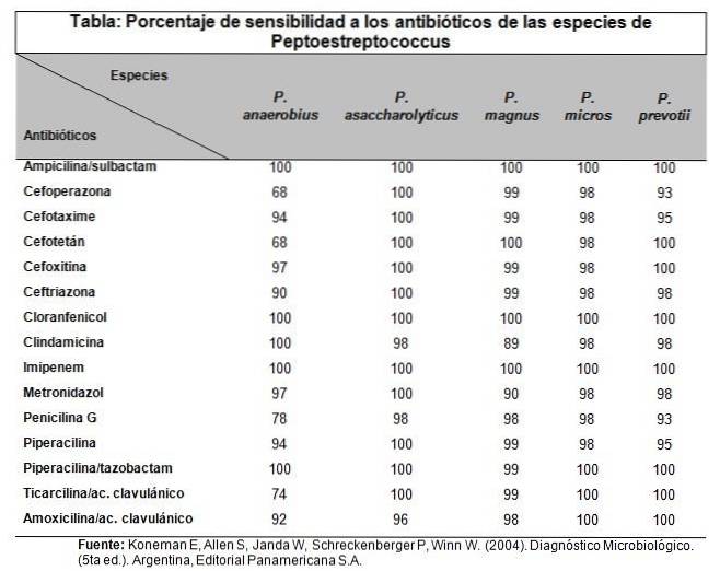
In general, the choice of antimicrobial is done empirically, since antimicrobial susceptibility methods are less standardized for slow-growing anaerobic bacteria.
Therefore, the approach is based on the expected susceptibility of anaerobes that commonly cause infections at the site in question..
Below is a table with detailed information on useful antibiotics.
In the case of infections caused by Pepto-streptococcus invasion of the oral microbiota to sterile sites, the way to prevent it is through good oral hygiene, which prevents the installation of gingival or periodontal diseases..
These injuries are usually the main source of entry. In the case of traumatic dental extractions, antibiotic therapy should be indicated to avoid infectious complications due to these microorganisms..
Likewise, when surgical or invasive procedures are practiced that may disrupt the state of any mucosa.


Yet No Comments