
The pyloroplasty is a surgical procedure that involves relaxing the pyloric sphincter muscle to prevent the reflux of acids and food from the duodenum into the stomach.
The pylorus is a ring-shaped muscle found in the last portion of the stomach and separates it from the duodenum. Its function is to allow and regulate the passage of stomach content into the duodenum and small intestine..
When the muscle that makes up the pyloric sphincter widens, the condition known as pyloric stenosis occurs. During the same the union channel between the stomach and the duodenum is obstructed, therefore there is a reflux of stomach contents (food and gastric acids). This disease can bring complications such as stomach ulcers and malnutrition.
In many cases, pyloroplasty is combined with another procedure known as vagotomy, in which the vagus nerve is cut to avoid hyper secretion of gastric acids in the stomach and duodenum..
Article index
Pyloroplasty is the surgical procedure performed to relax the pyloric sphincter and release its lumen..
Whether the muscle is enlarged and thick or there is an obstruction due to an ulcer, pyloroplasty is the type of surgery that is carried out to improve the patient's condition..
It is an abdominal surgery that consists of sectioning the pyloric sphincter muscle, achieving its relaxation and allowing food to pass back into the duodenum. It can be done by open approach or laparoscopically.

To avoid hyperactive acid secretion into the stomach and duodenal lumen, it is almost always combined with a treatment called vagotomy, in which the vagus nerve, which is responsible for stimulating gastric cells, is cut..
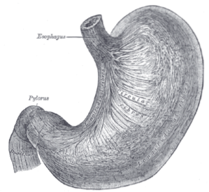
The stomach is a muscular organ of the digestive system that is responsible for storing and beginning the digestion of ingested food. Later, these foods are emptied into the duodenum to continue the digestion process..
It is located in the upper left part of the abdomen, being the continuation of the esophagus, which is the muscular channel of passage that joins it with the mouth.
It consists of two sphincters, one upper and one lower. The upper sphincter prevents the reflux of food and acids into the esophagus. It is known as cardia.
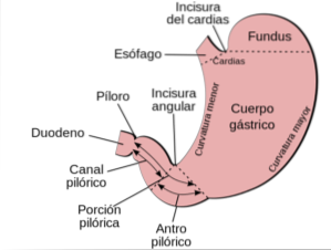
The lower sphincter separates it from the duodenum and regulates the emptying of gastric contents into the small intestine. It is called the pylorus.
The stomach has two parts, the fundus and the body. The fundus is located immediately after the cardia, a dome-shaped area that is in contact with the left diaphragm.
Just after the fundus is the body of the stomach, which is the largest portion of the organ and from where emptying occurs by a process mediated by the pylorus..
Within the body of the stomach, the process of chemical digestion occurs, which is when food mixes with stomach acids and other enzymes to break down and pass into the duodenum to continue its digestion..
The vagus nerve is responsible for providing motor and sensory innervation to the stomach. It has fibers that modulate the acid secretion process of stomach cells.
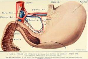
When there is food in the stomach, the vagus nerve activates the production and exit of gastric juices towards the stomach lumen and begins the mixing movement for the formation of the food bolus..
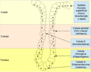
The stomach is an organ that serves to store and empty food into the duodenum. This is where an important step for digestion occurs, which is the mixing and decomposition of food by acids or gastric juices..
Gastric juices are a mixture of substances secreted by the stomach lining, it is composed mainly of hydrochloric acid, mucus, sodium and potassium chloride, bicarbonate and the enzyme pepsin.
The movements made by the stomach, in intervals of 20 minutes, mix the gastric juices with the food and form the chyme or food bolus. Chyme is an acid paste that passes into the duodenum in small amounts with each movement.
The passage of the chyme to the duodenum occurs through the periodic opening and closing of the pyloric sphincter. The complete passage of the bolus from the stomach to the duodenum takes approximately 4 hours.
The stomach does not perform nutrient absorption activity but it does prepare the food bolus with enzymes that break down carbohydrates and proteins so that these elements are absorbed in the duodenum and the rest of the small intestine..
There are substances that are absorbed in the stomach such as coffee, aspirin, alcohol and some vitamins.
In addition to these physiological functions, the gastric fundus is responsible for secreting the hormone ghrelin, called the hunger hormone. The secretion of this hormone sends impulses that indicate if the stomach is not distended and needs food.
The processes of acid secretion in the stomach occur in a balanced way. As food enters, the mechanism by which cells secrete acid into the stomach cavity is triggered..
In some cases there is an imbalance in this phase, with more acid than necessary. Therefore, the stomach mucosa and the duodenal mucosa end up subjected to an excessively acidic environment..
Some of the most common factors that trigger increased stomach acid secretion are frequent use of aspirin and infection with Helycobacter pyllori that causes great cellular damage.
Continuous increased secretion of gastric juice leads to the formation of gastric and duodenal ulcers. Gastroduodenal ulcers are wounds that originate in the mucosa of the stomach or duodenum due to continuous exposure of the mucosa to the acidic environment of the stomach..
The most common sites for ulcers are in the lesser curvature of the stomach, at the entrance to the pylorus, and in the duodenum. The diagnosis of ulcer is made through the study known as upper digestive endoscopy..
In upper digestive endoscopy, a special camera is introduced through the mouth into the duodenum to observe the state of the mucosa and take a biopsy if necessary..
Acute ulcers are swollen and sometimes bleeding wounds. Chronic ulcers have more scarred edges and are sometimes deep.
One of the complications of ulcers is obstruction. This means that a chronic ulcer has so much inflammation and generates such a large fibrosis around it that it ends up obstructing the lumen. It is a complication that can be seen in adults with gastro-duodenal ulcer disease. The most common is that there is obstruction of the pylorus or duodenum.
Another common cause of pyloric obstruction in young children, between 2 days and 3 weeks old, is pyloric hypertrophy. A condition in which the pyloric sphincter muscle is more developed than normal. This disease is characterized by low weight of the child, constant hunger, vomiting after eating and dehydration.

Yet No Comments