
The Peyer's patches They are anatomical regions located under the mucosa of the gastrointestinal tract, specifically in the lamina propria of the small intestine. They are sites of aggregation of a large number of lymphocytes and other accessory cells, which is why they represent part of the mucosal immune system.
Like the tonsils in the pharynx and the lymphoid follicles in the submucosa of the appendix, Peyer's patches resemble lymph nodes with respect to their structure and function, with the difference that the former are not encapsulated like the nodes..
It is important to remember that the immune response (the body's defense system against external "invaders") is mediated by various types of cells, lymphocytes being the most important, since, thanks to their ability to recognize antigens, they are responsible for triggering specific immune responses.
Peyer's patches were described in 1645 as "lymphoid follicles" by the Italian Marco Aurelio Severino, but it was not until 1677 that the term "Peyer's patches" was coined in honor of the Swiss pathologist Johann Conrad Peyer, who made a description detailed of them.
Its function, however, was determined many years later when, in 1922, Kenzaburo Kumagai noticed the ability to “absorb” pathogenic and foreign cells from the epithelium to the epithelial “dome” of Peyer's patches..
Article index
Peyer's patches belong to what is known as the “gut-associated lymphoid tissue” or GALT. Gut-TOassociated Lymphoid Tissue "), which is composed of lymphoid follicles distributed along the gastrointestinal tract.
This lymphoid tissue associated with the intestine represents one of the largest lymphoid organs in the body, since it contains almost 70% of the immune cells or "immunocytes".
A lymphoid follicle is an aggregate or set of lymphoid cells that does not have a defined structure or a particular organization..
Usually in gut-associated lymphatic tissue, these follicles are isolated from each other, but follicles in the ileum (the last portion of the small intestine) clump together to form Peyer's patches..
In the human small intestine, Peyer's patches are "oval" in shape and unevenly distributed. Cornes, in 1965, determined that the number of plaques during human development peaks between 15 and 25 years and subsequently decreases with age..
Other researchers have ensured that the area occupied by Peyer's patches in the ileum peaks during the third decade of life and that the largest proportion of these is concentrated in the last 25 cm of the ileum..
Like many other tissues in the human body, the organogenesis of Peyer's patches depends, to a large extent, on the participation of specific cytokines that mediate the differentiation and arrangement of these anatomical regions..
The main function of Peyer's patches as part of the immune system of the intestinal mucosa is to protect the "shell" of the intestines from invasion by potentially pathogenic microorganisms..
Some of the cells of the lymphoid follicles present in this "region" of the intestine are responsible for discriminating between pathogenic microorganisms and "commensalists" (which belong to the native microflora), since these follicles interact directly with the intestinal epithelium.
Peyer's patches participate in the “uptake” of foreign or pathogenic cells, however, it has been shown that cells belonging to this region are also capable of distinguishing between certain antigens and between non-pathogenic bacteria associated with the intestinal tract..
This process of recognition of the non-pathogenic is known as “oral tolerance” and it is an active process that leads to the formation of specific T lymphocytes that are capable of avoiding the triggering of an unnecessary immune response..
Oral tolerance is also defined as the antigen-specific elimination of humoral and cellular immune responses towards antigens that reach the body through the oral route, being especially useful for the protection of the intestinal mucosa against unfavorable inflammatory immune responses..
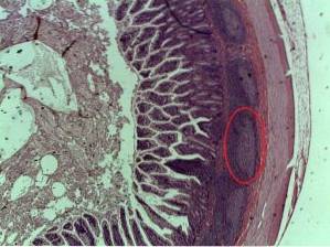
Peyer's patches are part of the lamina propria of the small intestine. The lamina propria is made up of loose connective tissue that, at the same time, is part of what is called the “nucleus” of the intestinal villi.
Various types of plasma cells, lymphocytes, leukocytes, fibroblasts, mast cells, and others are found in the lamina propria, and Peyer's patches are the portion of the lamina propria where permanent sets of lymphoid nodules or follicles are found..
Peyer's patches are architecturally distinguished into three main domains known as:
1- The follicular area
2- The interfollicular area and
3- The epithelium associated with the lymphoid follicles.
This region is comprised of lymphoid nodules or follicles characteristic of Peyer's patches that are composed of B cells (B lymphocytes) surrounded by a less compact (loose) portion of T cells (T lymphocytes) and many follicular dendritic cells or " antigen presenting cells ”(APC) TOntigen Presenting Cells).
The portion where lymphocytes or replicative B cells, dendritic cells, and another type of cell, macrophages, are found is called the "germinal center." Each lymphoid follicle, in turn, is surrounded by what is known as a “crown” or “subepithelial dome”.
The subepithelial dome also has a mixture of lymphoid cells (B and T lymphocytes), follicular dendritic cells and macrophages, and this is what the interfollicular area represents..
It has been shown that, in the lymphoid follicles of adult mice, the proportion of B cells in the internal region of these structures is more or less 50 or 70%, while the T cells represent only 10 to 30%.
Some research also suggests the presence of another specialized type of cells known as eosinophils, the proportion of which increases after exposure to oral allergens..
The ileum is lined by a simple epithelium (a single layer of cells) arranged cylindrically. However, in the regions adjacent to the lymphoid follicles of Peyer's patches there are a large number of squamous cells known as M cells, micro-fold cells or specialized membrane cells..
Apparently, the main function of M cells adjacent to these follicles is to capture antigens and direct or transfer them to macrophages that are also associated with Peyer's patches..
M cells do not have microvilli and are actively conducting pinocytosis to achieve transport from the lumen of the small intestine to the subepithelial tissues..
The mucosal-associated immune system is connected to the rest of the body's immune system thanks to the activation and migration capacity of T lymphocytes from Peyer's patches, which can reach the systemic circulation to exercise their immune functions..
Unlike the case of the epithelium of the mucosa of the intestinal villi, the epithelium associated with the lymphoid follicles has a low production of mucus, in addition, digestive enzymes are poorly expressed and the glycosylation patterns of the elements associated with glycocalyx are different.
Unlike other lymphoid tissues, such as lymph nodes, Peyer's patches do not have afferent lymphatic vessels that carry lymph "inside." However, they do have efferent drainage or efferent lymphatic vessels, capable of transporting lymph out of the lymphoid follicles.
The cells within the plaques are supplied by arterioles or small blood vessels capable of forming a capillary bed drained by high endothelial venules..
Given the important role played by Peyer's patches in the human body, there are a large number of associated pathologies, among which mention can be made of the following:
It is an inflammatory pathology characterized by recurrent inflammation of the digestive tract. The implication of Peyer's patches in this disease is due to the fact that the typical lesions of this cause the triggering of adaptive or innate immune responses to the bacterial flora.
In addition, it seems that Crohn's disease especially affects the distal portion of the ileum, right where there is a copious amount of Peyer's patches..
This condition is evident as a "battle" between grafts or transplants from one patient to another genetically incompatible..
The interaction between bacterial flora and the epithelial immune response is thought to contribute to the elicitation of inflammatory signals that contribute to stimulation of donor-derived T cells, mediated by host antigen-presenting cells..
The participation of Peyer's patches in this process was recognized by Murai et al., Who demonstrated that these structures are the anatomical site where infiltration of donor T cells occurs and where “anti-host” cytotoxic T cells are formed..


Yet No Comments