
The hodge blueprints are four imaginary anatomical divisions, which project onto the pelvis of the pregnant woman and serve as a guide to know the position of the fetus in the birth canal.
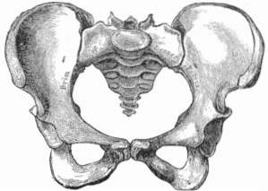
The pelvis is an anatomical cavity made up of the bones below the trunk. This cavity contains the internal reproductive organs. To divide the pelvis according to the Hodge planes, its anatomy must be well known.
The four planes are parallel imaginary lines and are drawn from specific points in the bones that make up the pelvis..
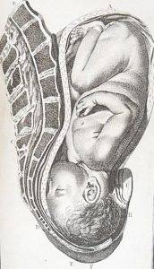
During labor, the fetus is descending from the pelvis to its exit through the vagina. Knowing the anatomical points of the pelvis to calculate the Hodge planes, it is possible to know how high the fetus is in the pelvis, in relation to the vaginal orifice.
By calculating the Hodge planes during a woman's labor, the doctor can tell if the labor is progressing normally or if, on the contrary, the labor is not being effective and other measures should be used to help expulsion of the fetus.
Article index
During pregnancy, the female pelvis undergoes various modifications, especially at the end of pregnancy. These changes become more important at the time of birth, when the fetus passes through the birth canal to be finally expelled..
The distance between the fetal head and the vaginal opening is known as the fetal height. A quick and efficient way to know this measurement is through the Hodge plans.
The pelvis is the bony structure under the spine, resting on the proximal bones of the legs (femur). These bones form a cavity where some abdominal organs and internal reproductive organs are located..
It is funnel-shaped, presenting a wide upper circumference known as the upper narrow and a lower circumference with a smaller diameter known as the lower narrow..
There are three bones that make up the pelvis: behind is the sacral bone, which is the terminal portion of the vertebral column, and in front the two iliac bones united in the pubis.
Hodge's planes are 4 parallel and imaginary lines that are numbered from top to bottom and that locate the height of the fetus with respect to the vagina at the time of delivery.
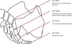
The planes are identified from the anatomical points of the pelvis as follows:
- Foreground: is the line that is drawn from the symphysis pubis to the junction of the fifth lumbar vertebra with the sacrum. This junction is also known as a promontory. Hodge's close-up coincides with the upper pelvis.
- Background: its identification is carried out by the union of the second sacral vertebra to the lower edge of the symphysis pubis.
- Third Plane: in this case, the line is drawn at the height of the ischial spines, parallel to the previous one. The ischial spines are two prominences of the lateral bones of the pelvis.
- Fourth Plan: for the last of the parallels, the union of the sacrum with the coccyx, known as the sacral vertex, is taken as a reference. From there a line is drawn parallel to all the previous ones.
When the fetus reaches this plane, it is about to be expelled.
Hodge planes are used in obstetric medical history. That is, the measurement is made exclusively in pregnant women.
When the doctor refers to the fetus based on the Hodge planes, he is giving a specific measurement of the fetal height.
Since the identification of the planes is known and used worldwide, these terms are universally understood without the need to provide more detailed data regarding the height at which the fetus is in the birth canal..
The identification of the Hodge planes is especially important during the passage of the fetus through the birth canal.
When taking the medical history of a woman in labor, the height at which the fetus is located is established through vaginal examination thanks to the Hodge planes. That way it's easy to keep track as the hours go by.
Once the anatomical points for the calculation of the imaginary parallels of Hodge are known, through the physical examination, the doctor can know how high the fetus is.
In a normal pregnancy, the fetus descends through the different pelvic planes. By means of vaginal examination, which is a test that the doctor performs by inserting two fingers through the vagina, the head of the fetus can be touched when it reaches the last two planes of Hodge.
Hodge's third and fourth planes correspond to the so-called fetal nesting. This means that the head of the fetus is completely insinuated into the birth canal and that the fetus is about to be expelled..
When a fetus does not progress beyond one of the planes, the patient must be studied to be able to make an adequate diagnosis and try to solve it by continuing the delivery.
In some cases, the diameter of the woman's pelvis is smaller than the head or shoulders of the fetus. For this reason, there can be a good progression between Hodge's first two shots, which are wider, and stop at the third, when the pelvis narrows..
If the fetus cannot progress further through the pelvis, it is vitally important to attend the delivery to avoid fetal distress..
The fetus manages to be born once the doctor manages to pass through the last bony area of the birth canal, corresponding to the fourth plane of Hodge. After exceeding this diameter, a vaginal delivery is achieved.
1. Bottle J; Clavero, J. (1993). Obstetric examination. Treaty of Gynecology.
2. Bonilla-Musoles, F; Pellicer, A. (2007). The canal and the object of childbirth. Basic Obstetrics, Reproduction and Gynecology.
3. Sicuranza, BJ; Tisdall, H; They read WM; Palmeri T. (1970). The planes of Hodge as an index of the progress of labor. The Journal of Reproductive Medicine. Taken from: ncbi.com
4. Steer, P; Flint, C. (1999). ABC of labor care: Physiology and management of normal labor. BMJ. Taken from: ncbi.com
5. Carvajal, H; Chambi, G. (2012). Anatomical description of the obstetric pelvis and pelvimetric examination in pregnant women. Bolivian Archives of Medicine. Taken from: scielo.isciii.es


Yet No Comments