The choroid plexuses They are small vascular structures of the brain. These regions are responsible for forming the cerebrospinal fluid, which is an essential element for the protection of the central nervous system..
Most of the cerebrospinal fluid originates from the choroid plexuses, renewing itself six to seven times a day in human brains. These structures stand out for being a continuation of the pia mater at the level of the ventricles. For this reason, these structures are formed mainly by modified epindymal cells..
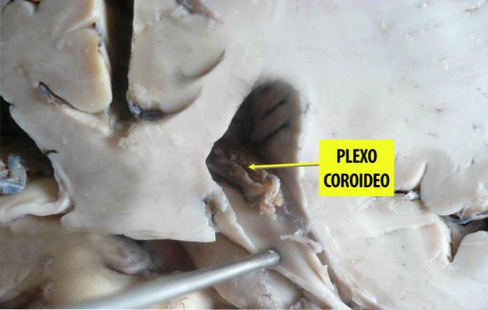
The choroid plexuses are a small region of the brain that is responsible for forming the cerebrospinal fluid, an intracranial substance that runs through different regions of the brain in order to provide protection.
Article index
More specifically, these elements of the brain constitute vascular structures that are located on the sides of the cerebral ventricles. They are regions formed by a large number of capillaries that constitute a network and are surrounded by cells with a structure similar to an epithelium..
In this sense, the choroid plexuses lack a basal lamina and have a sharp base with extensions that join the oligondrocytes to be able to use blood plasma, which is necessary to generate cerebrospinal fluid.
Together with the ependymal cells, these structures constitute a continuation of the pia mater (the internal meninx that protects the central nervous system) at the level of the ventricles..
Thus, the pia mater fulfills the same function as the choroid plexus. However, the first is carried out in the brain and spinal cord, while the second is located in the cerebral ventricles..
The human brain has four different choroid plexuses. Each of them is located in one of the four brain ventricles.
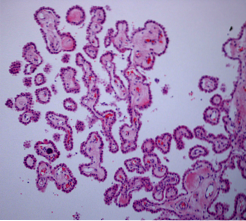
The choroid plexuses are formed by a layer of cuboidal epithelial cells that surround the nucleus of the capillaries and the connective tissue. The epithelial layer of the plexuses is continuous with the layer of ependymal cells, which covers the cerebral ventricles.
However, the ependymal cell layer, unlike the choroid plexuses, has a series of very tight junctions between cells. This fact prevents most substances from passing through the layer and reaching the cerebrospinal fluid..
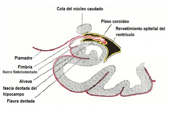
Regarding their location, the choroid plexuses are found in the upper region of the lower horn of the lateral ventricles..
They have a long structure that runs across the entire surface of the ventricle. Likewise, the choroid plexuses pass through the interventricular foramen and are present in the upper part of the third ventricle..
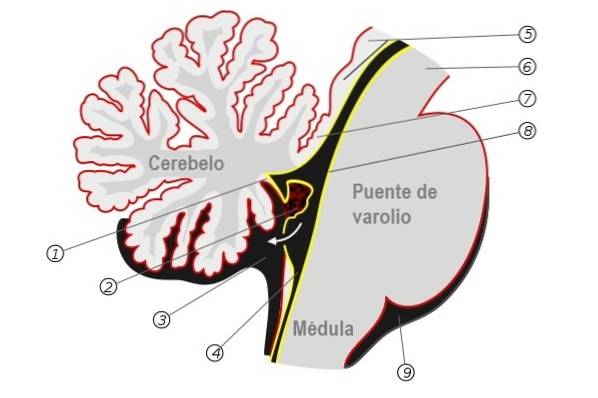
Furthermore, these structures can also be seen in the fourth cerebral ventricle. In this case, they are located in the section closest to the lower half of the cerebellum..
Thus, the choroid plexus constitutes a structure that is present in all components of the ventricular system except the cerebral aqueduct, the frontal horn of the lateral ventricle, and the occipital horn of the lateral ventricle..
The choroid plexuses configure the continuation of the pia mater at the level of the ventricles, they are formed by modified ependymal cells that have a basal lamina.
The cells of these plexuses are connected to each other through occlusive junctions, and they settle on the connective (non-nervous) tissue of the brain..
The ependymal cells of the choroid plexuses rest on the connective tissue and form a substance known as the choroidal tissue. This fabric folds to form the choroid plexuses, which are characterized by having a large number of capillaries immersed in their tissue..
The plasma from these capillaries is filtered through the epithelium of the choroid plexuses and acts as a dialyzing membrane. Finally, the plasma is sent to the ventricles as cerebrospinal fluid..
The main function of the choroid plexuses is to produce and transmit cerebrospinal fluid
Cerebrospinal fluid is a colorless substance that bathes the brain and spinal cord. It travels through the subarchnoid space, the cerebral ventricles and the ependymal canal, and has a volume of approximately 150 milliliters..
The main function of this substance is to protect the brain. Specifically, it carries out the following activities:
Beyond the production of cerebrospinal fluid, the choroid plexuses act as a filtration system, removing metabolic waste, foreign substances, and excess neurotransmitters in the cerebrospinal fluid..
Thus, these plexuses play a very important role in adapting and maintaining the extracellular environment that the brain requires to function properly..
At present, the main pathology related to the choroid plexuses is tumors. Specifically, three main types have been described: choroid plexus papilloma, atypical papilloma, and carcinoma..
These alterations are quite rare primary brain tumors in the general population. They are derived from the epithelium of the choroid plexus and are especially prevalent during childhood.
The location of these pathologies is, in most cases, the lateral ventricles. However, they can also originate in the fourth and third ventricle.
Its most common clinical presentation is hydrocephalus. Likewise, it can cause leptomeningeal dissemination in cases of papilloma and carcinoma..
Overall, choroid plexus tumors represent between 0.3% and 0.6% of all brain tumors. Of the three types, papillomas are much more frequent, while carcinomas have a very low prevalence.


Yet No Comments