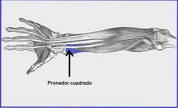
The pronator square or musculus pronator quadratus It is a muscle that is located at the level of the wrist. It is the main person in charge of activating the pronation movement of the forearm..
The term pronator comes from the Latin word pronation, which means rotation. Its name refers to its function. This muscle works in conjunction with the pronator teres muscle..
Their joint action makes possible the rotational movement of the forearm and the hand. The movement consists of rotating the forearm in such a way that the back of the hand is facing upwards. This movement is called pronation. The opposite movement is called supination.
This muscle is innervated by a branch of the median nerve called the anterior interosseous nerve, which is very vulnerable to compression and traction due to its location and trajectory..
The pronator quadratus muscle is very powerful and with a small contraction of the muscle it is capable of producing movement. So much so that patients who have suffered a double diaphyseal fracture of the upper limb (ulna and radius at the same time), have difficulty fully recovering from pronation and supination movements.
Usually there is a defective union of the bones with pseudarthrosis. This is very common and is due to the angulatory force exerted by the biceps and pronator quadratic muscles on the bones in recovery -even when trying to be at rest-, preventing correct rotational alignment..
Article index
The pronator square muscle has a quadrilateral shape, being thin and flattened in appearance..
It is closely related to the flexor tendons of the wrist, as it is the deepest muscle in the region. It connects anteriorly with the deep flexor, the great palmar, the great flexor of the thumb, as well as the ulnar anterior and the ulnar and radial arteries.
While on the posterior side it has a connection with the radius, the inter-bone ligament and the ulna..
At its ends (insertion part) it has an aponeurotic texture, that is, fibrous, while the rest of the muscle is fleshy.
This consists of two heads, a superficial and a deep one. Both originate in the anterior distal area of the ulnar shaft, but the superficial one attaches to the shaft of the radius, while the deep one implants into the proximal ulnar notch..
The fibers of the pronator quadratus muscle are oriented perpendicular to the direction of the forearm.
The pronator quadratus muscle can atrophy in the practice of certain sports that require repetitive and persistent movement (forearm rotation) or other activities that lead to the contraction of the pronator muscles, both the round and the square.
The pronator quadratus comes out of the anterior and inferior portion of the ulna bone.
The pronator quadratus muscle inserts at the level of the distal quarter of the external portion of the radius.
It is innervated by the interosseous branch that comes from the median nerve.
The pronator quadratus muscle helps hold the ulna and radius bones together.
On the other hand, together with the pronator teres, it allows pronation of the proximal radioulnar and humerus-radial joints (elbow), which contributes to the pronation of the hand and forearm (the ulna and the radius overlap forming an X ). The pronation movement originates from supination (starting position).
It is a muscle that presents great power. With a slight contraction it already generates movement.
Among the congenital anomalies that can be found are: the muscle may be absent or found divided into two different bellies.
It is also known as Kiloh-Nevin syndrome, in honor of the doctors who described the disease in 1952. It is characterized by compression of the interosseous nerve.
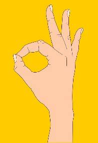
This clinical entity should be suspected when the patient reports pain in the anterior region of the forearm, inability to flex the thumb joint and difficulty in performing certain activities, such as: writing or expressing the "ok" sign with the thumb. and index.
To confirm the diagnosis, an MRI or electromyography may be requested..
León et al. Proposed a surgical technique for the treatment of scaphoid nonunion, through the pedunculated pronator square bone graft. In all operated cases they obtained satisfactory results.
The pronator quadratus, being a muscle with a very deep location, cannot be palpated.
To evaluate the function of both pronators (square and round), the patient is asked to turn the palm of the hand downwards and try to hold, while the examiner tries to turn the hand upwards, until reaching complete supination. If there is pain the test is positive.
This exercise comprises the complete pronation and supination movement, it is generally used in physiotherapeutic consultations to evaluate the recovery of movement of patients who have suffered from paralyzing disease, fractures or muscle hypertrophy, among others..
The patient is positioned seated with the forearm flexed at a 90 ° angle to the arm. The starting position will be with the hand laterally. Subsequently, the patient is asked to turn the hand in such a way that the back of the hand is facing downwards (pronation movement).
Then you are asked to do the opposite from the starting position (supination movement). With this simple exercise, several muscles are exercised.
In the pronation movement: the round and square pronator muscle.
In the supination movement: biceps brachii muscle, short supinator muscle and long supinator muscle.
If the exercise is carried out without problem, or pain and in the same way with both extremities (right and left) the muscles mentioned are in good condition.

Yet No Comments